. 2025 Oct 1;18(10):dmm052123.
doi: 10.1242/dmm.052123. Epub 2025 Sep 18.

Generation of human induced pluripotent stem cell lines from patients with FGFR2-linked syndromic craniosynostosis

Affiliations

Generation of human induced pluripotent stem cell lines from patients with FGFR2-linked syndromic craniosynostosis

Max Gijsbertsen et al. Dis Model Mech. .

Abstract

Craniosynostosis is a multigenic congenital condition in which one or more calvarial sutures have prematurely fused during the development of the fetus. Pathogenic variants in FGFR2 are associated with the development of syndromic craniosynostosis, such as Crouzon, Apert and Pfeifer syndromes. Investigation of FGFR2-linked craniosynostosis is hindered by the lack of appropriate in vitro models. Patient-derived human induced pluripotent stem cell (hiPSC) in vitro disease models provide the opportunity to investigate the disease, identify molecular targets for pharmaceutical treatments, and enable the generation of autologous pluripotent stem cell catalogues. Here, we report three patient-derived hiPSC lines carrying the C342Y, S252W or E565G FGFR2 pathogenic variant. The patient hiPSC lines express characteristic pluripotency markers and display distinct phosphorylation profiles under unstimulated conditions. FGFR2C342Y showed autophosphorylation in the absence of bFGF ligand, although downstream docking proteins PLCγ and FRS2α were not phosphorylated. FGFR2S252W and FGFR2E565G hiPSCs showed increased phosphorylation of docking proteins PLCγ and FRS2α, whereas FGFR2 was not phosphorylated. These patient hiPSC lines provide molecular and cellular options to investigate FGFR2-linked craniosynostosis in the patient-specific genomic context and develop therapeutic modalities.

Keywords: Apert; Craniosynostosis; Crouzon; FGFR2; Pfeiffer; hiPSC.


Conflict of interest statement

Competing interests The authors declare no competing or financial interests.

Figures
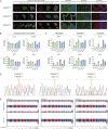
Fig. 1.
Fig. 1.
Identification of pluripotency in patient-derived human induced pluripotent stem cell (hiPSC) lines FGFR2C342Y, FGFR2S252W and FGFR2E565G. (A) Immunocytochemistry for embryonic stem cell markers SSEA4, NANOG, TRA-1-81 and OCT-4 (left panel), and trilineage markers NCAM, SOX17 and β-tubulin (right panel), in patient-derived hiPSCs. (B,C) RT-qPCR analysis of trilineage and embryonic stem cell markers of patient-derived hiPSCs. The mRNA expression of embryonic stem cell markers (B) and trilineage markers (C) of multiple human embryonic stem cell (HuES) clones (green bars) are used as reference values for the determination of expressed RNA markers in the reported hiPSC lines FGFR2C342Y, FGFR2S252W and FGFR2E565G (blue bars). Gene of interest values are corrected against GAPDH reference gene expression. Error bars display mean±s.d. Replicates used in each cell line tested n=3. (D) Identification of the respective craniosynostosis variant and genomic stability of the patient-derived hiPSCs. The orange bars highlight the nucleotide location of the patient pathogenic variant. (E) Compressed genomic overview of Global Screening Array analysis of the three patient-derived hiPSC lines FGFR2C342Y, FGFR2S252W and FGFR2E565G. B-allele frequency and Log(R) ratio are listed on the y-axis. Chromosome numbers are listed on the x-axis and in the graph in alternating blue and red coloration.
Fig. 2.
Fig. 2.
Protein and phosphorylation analysis of FGFR2 and docking proteins PLCγ and FRS2α in bFGF-free conditions. (A) Western blot chemiluminescence images of healthy control samples 1 and 2, and patient samples FGFR2C342Y, FGFR2S252W and FGFR2E565G. For each sample and condition, 10 µg protein-isolated whole-cell lysate is loaded. The red arrowhead indicates the secondary lower-molecular mass band of FGFR2. Technical triplicates (n=3) are derived from three parallel, but separate, cultures. (B) Comparative quantification of protein analysis through western blotting of FGFR2, PLCγ, FRS2α and their phosphorylated forms in bFGF-free conditions. Data from healthy control samples 1 and 2 are shown as green bars. Data from patient samples FGFR2C342Y, FGFR2S252W and FGFR2E565G are shown as blue bars. Protein values are corrected against actin loading control values. Statistical analysis was performed using one-way ANOVA and Tukey's multiple comparison post-hoc test. Error bars display mean±s.d. Replicates used in each cell line tested n=3. Statistical significance is displayed in compact letter display format, with the significance threshold of P<0.05. (C) A table summarizing the observations on protein expression and phosphorylation of FGFR2, PLCγ and FRS2α for all three patient lines. Equal marks represent no significant difference compared to healthy controls. Upward- and downward-pointing arrows indicate significant upregulation or downregulation, respectively, compared to healthy control 1. (D) Schematic model speculating how the various patient FGFR2 pathogenic variants impact their phosphorylation and activation of the docking proteins in an unliganded state according to published literature (reviewed in Ornitz and Itoh, 2015) and our findings. WT, wild type. Created in BioRender by Gijsbertsen, M. (2025). https://BioRender.com/dhiibrf. This figure was sublicensed under CC-BY 4.0 terms.

References

    1. Ahmed, Z., Schüller, A. C., Suhling, K., Tregidgo, C. and Ladbury, J. E. (2008). Extracellular point mutations in FGFR2 elicit unexpected changes in intracellular signalling. Biochem. J. 413, 37-49. 10.1042/BJ20071594 - DOI - PubMed
    1. Ahmed, Z., George, R., Lin, C.-C., Suen, K. M., Levitt, J. A., Suhling, K. and Ladbury, J. E. (2010). Direct binding of Grb2 SH3 domain to FGFR2 regulates SHP2 function. Cell. Signal. 22, 23-33. 10.1016/j.cellsig.2009.08.011 - DOI - PubMed
    1. Al Abbar, A., Ngai, S. C., Nograles, N., Alhaji, S. Y. and Abdullah, S. (2020). Induced pluripotent stem cells: reprogramming platforms and applications in cell replacement therapy. BioRes. Open Access 9, 121-136. 10.1089/biores.2019.0046 - DOI - PMC - PubMed
    1. Anderson, J., Burns, H. D., Enriquez-Harris, P., Wilkie, A. O. and Heath, J. K. (1998). Apert syndrome mutations in fibroblast growth factor receptor 2 exhibit increased affinity for FGF ligand. Hum. Mol. Genet. 7, 1475-1483. 10.1093/hmg/7.9.1475 - DOI - PubMed
    1. Bergquist, C. S., Nauta, A. C., Selden, N. R. and Kuang, A. A. (2016). Age at the time of surgery and maintenance of head size in nonsyndromic sagittal craniosynostosis. Plast. Reconstr. Surg. 137, 1557-1565. 10.1097/PRS.0000000000002049 - DOI - PubMed

MeSH terms

Substances

LinkOut - more resources