Multicenter Study
. 2025 Dec;17(1):2545420.
doi: 10.1080/19490976.2025.2545420. Epub 2025 Aug 22.

Increased reflux secondary bile acids are associated with changes to the microbiome and transcriptome in Barrett's esophagus

Affiliations
Multicenter Study

Increased reflux secondary bile acids are associated with changes to the microbiome and transcriptome in Barrett's esophagus

Ceylan Tanes et al. Gut Microbes. 2025 Dec.

Abstract

Bile acids are a major component of gastro-esophageal refluxate, thought to contribute to the development of Barrett's esophagus (BE) and esophageal adenocarcinoma (EAC). As the microbiome shifts with EAC progression and bile acids influence bacterial composition, we examined these connections in a multi-center, cross-sectional study. We analyzed biospecimens from patients undergoing endoscopy using LC-MS to quantify bile acids in gastric aspirates, 16S rRNA sequencing for tissue microbiome profiling, and RNA sequencing on BE or cardia tissue. Among 153 patients (52 controls, 101 BE: 50 no dysplasia, 10 indefinite, 17 low-grade dysplasia, 17 high-grade dysplasia, and 7 EAC), we observed increased Streptococcus in BE tissue; dysplasia and EAC were associated with more Lactobacillus and decreased Actinomyces and other genera. Refluxate bile acids were mainly conjugated, indicating minimal bacterial metabolism, while BE patients had elevated secondary bile acid levels. Streptococcus correlated with upregulation of IL6, FGF2, and HGF, and decreased Actinomyces showed the most associations with gene expression, including the oxidative phosphorylation pathway. We identified two distinct BE gene expression clusters independent of histology, bile acid, or microbiome composition. These findings suggest bile acids shape the BE microbiome and associate with gene expression changes potentially relevant to EAC development.

Keywords: Barrett’s esophagus; bile acids; esophageal adenocarcinoma; microbiome.


Conflict of interest statement

No potential conflict of interest was reported by the author(s).

Figures
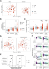
Figure 1.
Figure 1.
Patients with Barrett’s esophagus have a distinct tissue-associated microbiome. A) Principal coordinates analysis (PCoA) of weighted UniFrac distances show no evidence of significant clustering comparing BE with non-BE in saliva (left) and oral swash (right); B) the BE tissue microbiome was more closely related to oral wash within individuals as compared to controls off of PPIs (p < 0.001) and non-significantly compared to controls on PPIs (p = 0.07). Shown are within-individual weighted UniFrac distances compared to oral wash; C) the gram positive:negative ratio was higher in BE tissue compared to cardia in controls (vs. PPI users, p < 0.001; vs. PPI non-users, p < 0.001); D) PCoA of weighted UniFrac distances for squamous and BE/cardia tissue demonstrating significant differences in beta diversity comparing BE with controls (controlled for PPI use and antibiotics; squamous, PERMANOVA p = 0.003; BE/cardia, PERMANOVA p = 0.001); E) BE patients had significant differences in the relative abundance bacterial genera compared to controls in both squamous and BE tissue; F) bacterial genera with significantly increased and decreased relative abundance associated with stages of progression from BE to EAC. *p < 0.05, **p < 0.01, ***p < 0.001.
Figure 2.
Figure 2.
Secondary bile acid levels are increased in refluxate in patients with Barrett’s esophagus. A) virtually all of the measure bile acids were primary and secondary conjugated bile acids; B) patients with BE had significantly greater secondary bile acids compared to controls; C) there were no significant differences in bile acid composition across stages of BE to EAC; D) in BE/cardia tissue, there were non-significant trends towards positive correlations between bile acid levels and gram-positive bacteria and inverse correlations with gram-negative bacteria. The bile acid:bacteria correlations diminished with more proximal sampling sites.
Figure 3.
Figure 3.
Gene expression changes in patients with BE. A) unsupervised clustering analyses demonstrated three distinct clusters, one comprised of controls and two BE clusters independent of associated histology; (B) heatmap of 40 genes that were significantly differentially expressed in all pairwise comparisons between clusters. C) Plot demonstrating large number of differentially expressed genes in BE, including numerous markers of intestinalization; D) among patients with BE, LAIR2 was significantly increased and RPL10P9 significantly decreased across stages from BE to EAC (analyses adjusted for clinical risk factors for EAC). NDBE: non-dysplastic Barrett’s esophagus; IND: indefinite for dysplasia; LGD: low grade dysplasia; HGD: high grade dysplasia; EAC: esophageal adenocarcinoma. * p<0.05, ** p<0.01, ***p<0.001.
Figure 4.
Figure 4.
Relationships between the microbiome and bile acids with tissue gene expression. A) Volcano plots demonstrating significant associations between relative abundance of bacterial genera associated with BE progression to EAC and tissue gene expression; B) up- and down-regulated genes associated with the secondary bile acids GDCA and TDCA and with total conjugated secondary bile acids.

References

    1. Siegel RL, Miller KD, Wagle NS, Jemal A.. Cancer statistics, 2023. CA Cancer J Clin. 2023;73(1):17–14. doi: 10.3322/caac.21763. - DOI - PubMed
    1. Ward JBJ, Lajczak NK, Kelly OB, O’Dwyer AM, Giddam AK, Ní Gabhann J, Franco P, Tambuwala MM, Jefferies CA, Keely S, et al. Ursodeoxycholic acid and lithocholic acid exert anti-inflammatory actions in the colon. Am J Physiol Gastrointest Liver Physiol. 2017;312(6):G550–G558. doi: 10.1152/ajpgi.00256.2016. - DOI - PubMed
    1. Roberts DC, Chidambaram S, Kinross JM. The role of the colonic microbiota and bile acids in colorectal cancer. Curr Opin Gastroenterol. 2022;38(2):179–188. doi: 10.1097/MOG.0000000000000816. - DOI - PubMed
    1. Quante M, Bhagat G, Abrams JA, Marache F, Good P, Lee M, Lee Y, Friedman R, Asfaha S, Dubeykovskaya Z, et al. Bile acid and inflammation activate gastric cardia stem cells in a mouse model of Barrett-like metaplasia. Cancer Cell. 2012;21(1):36–51. doi: 10.1016/j.ccr.2011.12.004. - DOI - PMC - PubMed
    1. Tian Y, Gui W, Koo I, Smith PB, Allman EL, Nichols RG, Rimal B, Cai J, Liu Q, Patterson AD. The microbiome modulating activity of bile acids. Gut Microbes. 2020;11(4):979–996. doi: 10.1080/19490976.2020.1732268. - DOI - PMC - PubMed

Publication types

MeSH terms

LinkOut - more resources