Comparative Study
. 2025 Sep;11(5):e70594.
doi: 10.1002/vms3.70594.

Comparative Detection and Genetic Characterization of Feline Panleukopenia Virus in Bangladesh

Affiliations
Comparative Study

Comparative Detection and Genetic Characterization of Feline Panleukopenia Virus in Bangladesh

Nurejunnati Jeba et al. Vet Med Sci. 2025 Sep.

Abstract

Background: Feline panleukopenia virus (FPV) is a highly contagious and often fatal disease affecting domestic and wild felines. Accurate diagnosis and understanding of circulating strains are essential for effective control.

Objectives: This study aimed to evaluate the diagnostic accuracy of a rapid immunochromatographic (IC) antigen test compared to PCR for FPV detection in clinically suspected pet cats in Bangladesh. It also aimed to investigate the genetic and evolutionary characteristics of circulating FPV strains.

Methods: Faecal or rectal swab samples from suspected cats were tested using both IC strip tests and PCR. Sensitivity and specificity of the IC test were analysed using PCR as the reference. Partial sequencing of the VP2 gene was performed on four PCR-positive samples for phylogenetic and mutational analysis. Structural modelling of VP2 proteins was conducted to predict conformational changes.

Results: The IC test detected FPV in 84% of cases, whereas PCR confirmed only 60%, indicating a 24% false-positive rate. PCR showed higher diagnostic reliability. FPV prevalence was 92% among unvaccinated cats. Phylogenetic analysis of VP2 sequences revealed close genetic similarity with Chinese and Portuguese strains, suggesting possible cross-border transmission. Mutations such as A756G, A896G, E299G and T236I were consistently observed. Structural modelling indicated minor conformational changes in VP2.

Conclusion and clinical significance: PCR offers superior accuracy over IC testing for FPV diagnosis. Mutational changes may impact antigenicity and diagnostic performance. Improved diagnostic accuracy, molecular surveillance and updated vaccination strategies are essential to control FPV outbreaks in feline populations.

Keywords: Feline panleukopenia virus; IC strip test; PCR; VP2 gene mutations; phylogenetic analysis.


Conflict of interest statement

The authors declare no conflicts of interest.

Figures

FIGURE 1
FIGURE 1
Geographical representation of the locations included in this FPV study. The map was created using QGIS software.
FIGURE 2
FIGURE 2
Identification of FPV. (A) Preliminary detection using the IC test. Coloured lines in both the test (T) and control (C) regions indicate the presence of the target analyte in the sample, confirming a positive result. (B) Identification by PCR. Gel electrophoresis showing the VP2 gene amplicons of FPV (698 bp). Lanes: M—100 bp DNA ladder (Thermo Fisher, USA), N—Negative control, C—Positive control, Lanes 1–9—Representative FPV‐positive samples showing bands at approximately 698 bp. FPV, Feline panleukopenia virus.
FIGURE 3
FIGURE 3
Distribution of the cat population based on clinical signs and symptoms. The clustered column bar diagrams illustrate the number of FPV‐infected cats exhibiting clinical symptoms such as diarrhoea, fever, weakness, vomiting, anorexia and foul‐smelling faeces.
FIGURE 4
FIGURE 4
Distribution of FPV‐infected cats by age, sex and vaccination status. (A) Age distribution: cats aged ≤5 months versus >5 months. (B) Sex distribution: male versus female cats. (C) Vaccination status: vaccinated versus unvaccinated cats. (D) Breed distribution: Persian, local and mixed breeds.
FIGURE 5
FIGURE 5
Phylogenetic tree of FPV isolates identified in this study. (A) Neighbour‐Joining tree was constructed using 79 partial VP2 gene sequences retrieved from GenBank. Evolutionary distances were calculated using the Tamura–Nei method. Sequence alignment and evolutionary analyses were performed using MEGA11 software and ClustalW. Sequences obtained in this study are highlighted in blue.
FIGURE 6
FIGURE 6
Nucleotide substitution analysis of the VP2 gene in identified FPV isolates. Mutational analysis was conducted by aligning VP2 gene sequences from this study with reference sequences using CLC Sequence Viewer 8.0. Dots represent identical residues, whereas letters indicate nucleotide substitutions.
FIGURE 7
FIGURE 7
Amino acid variations in the VP2 protein of identified FPV isolates. Amino acid sequences of VP2 proteins were aligned and compared with reference strains. Dots indicate identical residues, whereas letters denote substitutions. Analysis was performed using CLC Sequence Viewer 8.0.
FIGURE 8
FIGURE 8
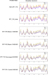
Secondary structure analysis of the VP2 protein in identified FPV isolates and reference strains. Secondary structures were predicted and analysed using the SOPMA online server with default parameters.
FIGURE 9
FIGURE 9
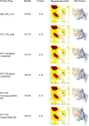
Prediction and analysis of the tertiary structure of the VP2 protein. The hydrophobicity of the predicted VP2 protein structures was analysed using SWISS‐MODEL and compared with reference proteins. Identity, Z‐score, Ramachandran plots and hydrophobic surface regions (orange patches) are depicted.
FIGURE 10
FIGURE 10
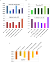
Physicochemical properties of the VP2 protein compared with reference proteins. Clustered column bar diagrams depict various properties. (A) Molecular weight and aliphatic index. (B) Theoretical pI and instability index. (C) GRAVY value of the VP2 capsid protein.

Similar articles

References

    1. Abdelbaky, M. M. , El‐Khabaz K. A., and Hamed M. I.. 2024. “Diagnostic Performance of a Rapid in‐Clinic Test for the Detection of Feline Parvovirus.” Assiut Veterinary Medical Journal 70, no. 182: 253–262.
    1. Alessa, M. , Marzok M., Elnafarawy H., et al. 2026. “Prevalence of Feline panleukopenia (FPL) in Domestic Cats: A Systematic Meta‐Analysis.” Veterinary Integrative Sciences 24, no. 1: 1–17. 10.12982/VIS.2026.001. - DOI
    1. Awad, R. A. , Khalil W. K. B., and Attallah A. G.. 2018. “Epidemiology and Diagnosis of Feline panleukopenia Virus in Egypt: Clinical and Molecular Diagnosis in Cats.” Veterinary World 11, no. 5: 578–584. 10.14202/vetworld.2018.578-584. - DOI - PMC - PubMed
    1. Balboni, A. , Bassi F., De Arcangeli S., et al. 2018. “Molecular Analysis of Carnivore Protoparvovirus Detected in White Blood Cells of Naturally Infected Cats.” BMC Veterinary Research 14, no. 1: 41. 10.1186/s12917-018-1356-9. - DOI - PMC - PubMed
    1. Baratloo, A. , Hosseini M., Negida A., and El Ashal G.. 2015. “Part 1: Simple Definition and Calculation of Accuracy, Sensitivity and Specificity.” Emergency 3, no. 2: 48–49. - PMC - PubMed

Publication types

MeSH terms

LinkOut - more resources