. 2025 Aug 22;20(8):e0330487.
doi: 10.1371/journal.pone.0330487. eCollection 2025.

Cardiac function and extracellular matrix morphology are altered by chronic high fat diet in Drosophila larvae

Affiliations

Cardiac function and extracellular matrix morphology are altered by chronic high fat diet in Drosophila larvae

Rachel M Andrews et al. PLoS One. .

Abstract

Cardiovascular disease is characterized by aberrant and excessive extracellular matrix (ECM) remodelling, termed fibrosis. Fibrotic remodelling is typically triggered by inflammation, which occurs systemically in obesity. Despite the contribution of fibrosis to adverse clinical outcomes and disease progression, there are no available treatments for this condition. Developing therapeutics for chronic conditions requires an understanding of in vivo ECM regulation, and how the ECM responds to a systemic challenge. We have therefore developed a Drosophila model for obesity via chronic high fat diet feeding of larvae and evaluated the response of the cardiac ECM to this metabolic challenge. We found that this model displays a striking reorganisation of the cardiac ECM, with fibres oriented anterior to posterior, rather than in a complex network, suggesting tension modulation is altered. We also observe corresponding deficits in heart function, with high fat diet treatments resulting in an inability to contract the heart effectively at systole. Our study reveals that different genotypes tolerate different levels of dietary fat, and that some genotypes may require a different dietary supplementation regime to generate a cardiac phenotype. In summary, the Drosophila model for chronic high fat diet recapitulates many of the defects observed in human cardiovascular disease, allowing further evaluation of genetic and environmental influences on cardiac structure and physiology in disease states.


Conflict of interest statement

The authors have declared that no competing interests exist.

Figures
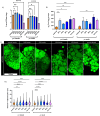
Fig 1
Fig 1. y1w1118 larvae fed a high fat diet display a dose dependent increase in markers of obesity.
Larval mass is not lowered by high fat diet supplementation, in contrast to high sucrose supplementation (A). HFD feeding results in a dose-dependent increase in triglyceride levels in y1w1118 larvae, especially females (B). Lipid droplets were visualized with BODIPY 493/503 (C) and both HFD feeding and a high sucrose diet result in a dose-dependent increase in lipid droplet size (D). Error bars in A and B are SEM. Scale bar in C is 100µm. White lines in D represent the median, dotted lines represent quartiles. n > 10 individuals for all groups. * = p < 0.05, ** = p < 0.01, *** = p < 0.001, **** = p < 0.0001. If no p value is indicated, comparison is not statistically significant.
Fig 2
Fig 2. vkgGFP larvae have elevated baseline triglyceride levels and do not tolerate HFD supplementation as well as y1w1118.
vkgGFP larval mass is lower in 20% and 30% HFD fed females and unchanged in other HFD supplementation groups (A). vkgGFP larvae did not have significantly elevated triglyceride levels in response to HFD feeding (B), but control individuals were found to possess markedly elevated baseline triglyceride levels compared to y1w1118 larvae (C). Larvae of the genotype Oregon R were found to have intermediate triglyceride levels to vkgGFP and y1w111 (C). HFD supplementation was not found to affect the viability of y1w1118 larvae (D), but did result in reduced viability of vkgGFP larvae (E). Error bars are SEM. * = p < 0.05, ** = p < 0.01, *** = p < 0.001, **** = p < 0.0001. If no p value is indicated, comparison is not statistically significant.
Fig 3
Fig 3. Pericardin fibre organization is perturbed in y1w1118 dietary treatments.
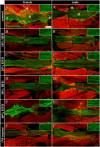
Controls demonstrate a normal, organized meshwork (A-A’) while dietary treatments exhibit a change in matrix organization, with matrix fibres becoming oriented anterior-posterior (B-F’). The cardiac ECM is visualized by immunolabelling Pericardin (green) and F-actin (red). The F-actin label in the background is body wall muscle. In panel A, H labels the heart tube, M labels alary muscles, a is anterior, p is posterior. All images are oriented anterior to the left. Pericardin insets are enlarged approximately 2.5x. Scale bar in A is 100µm. White line in panel B highlights the anterior-posterior orientation of Pericardin fibres. n > 10 for all groups.
Fig 4
Fig 4. Matrix organization reveals severe rearrangement in dietary treatments.
Matrix organization was scored on a scale of 1 (normal matrix organization) to 3 (the majority of matrix organization is disrupted). Dietary treatments demonstrate a linearity phenotype that is more common and more severe than controls in both y1w1118 (A) and vkgGFP larvae (B). In females, there is a downward trend in the size of the Pericardin matrix relative to the heart tube in y1w1118 (C) and vkgGFP (D). Males of all genotypes fail to reveal a significant trend. n > 10 for all groups. Panels A and B use a Chi squared test, figures C and D a one-way ANOVA with Dunnett’s correction. * = p < 0.05, ** = p < 0.01, **** = p < 0.0001. If no p value is indicated, comparison is not statistically significant.
Fig 5
Fig 5. Collagen-IV distribution is abnormal in vkgGFP dietary treatments.
In control individuals the Collagen-IV matrix possesses a uniform, sheet-like distribution across the surface of the heart (A’). HFD treated larvae exhibit elevated levels of clumping within the Collagen-IV matrix, indicated by white arrows, that is not found in the high sucrose diet treatment (B’, C’, D’, E’, F’). Gaps are observed in the high sucrose Collagen-IV matrix, indicated by white arrowheads (F’). The cardiac ECM is visualized by endogenous vkgGFP fluorescence (green), and immunolabelling of Pericardin (red) and F-actin (blue). In panel A, H labels the heart tube, M labels alary muscles, a is anterior, p is posterior. vkgGFP is also expressed in the trachea, indicated by T in panel B’. All images are oriented anterior to the left. Scale bar in A is 100µm. n > 10 for all groups.
Fig 6
Fig 6. Collagen-IV clumping depends on dosage of HFD treatment.
HFD feeding results in a dose-dependent increase in the amount of clumping within the Collagen-IV matrix (p < 0.0001) (A). High sucrose supplementation did not demonstrate increased Collagen-IV clumping compared to controls while the calorically comparable HFD does (CI = control −0.190896–1.5403, sucrose 1.13279–2.864116, 20% HFD 3.4414–5.131001) (B). The Collagen-IV matrix does not possess more gaps with HFD supplementation (p > 0.05) (C). High sucrose supplementation results in a Collagen-IV matrix with more gaps when compared to both controls diets and the calorically equivalent HFD treatment (CI = control 0.153–0.673, sucrose 0.974–1.494, 20% HFD 0.396–0.904) (D). Graphs in A and C are a model estimate, shown by the solid line, with 95% confidence intervals, indicated by the shaded area. In both A and C 0% supplementation refers to the control diet. Error bars in B and D represent 95% confidence intervals. n > 10 for all groups.
Fig 7
Fig 7. Optical coherence tomography reveals impaired contraction with HFD treatment.
OCT was used to visualize the heart beating in cross-section, revealing the area inside the lumen at both diastole and systole (A). Control hearts are round or oval and contract evenly along the perimeter, while HFD treatment hearts are abnormally shaped and unable to contract evenly on all sides (B). Lumen cross-sectional area in y1w1118 females displays a change in diastole only in high sucrose individuals (C), but does reveal increases in systolic area at higher concentrations of HFD. A similar trend is observed in males (D). HFDs demonstrate a dose-dependent impairment of heart contractions (E). Yellow outlines in A and B trace the heart lumen. Yellow arrows in B indicate plane of contraction. Error bars in C and D are SEM. Graph in E is a model estimate, shown by the solid line, with 95% confidence intervals, indicated by the shaded areas. n > 10 for all groups. * = p < 0.05, ** = p < 0.01, *** = p < 0.001, **** = p < 0.0001. In panels C and D, if no p value is indicated the comparison was not statistically significant.

References

    1. Sidney S, Lee C, Liu J, Khan SS, Lloyd-Jones DM, Rana JS. Age-Adjusted Mortality Rates and Age and Risk-Associated Contributions to Change in Heart Disease and Stroke Mortality, 2011-2019 and 2019-2020. JAMA Netw Open. 2022;5(3):e223872. doi: 10.1001/jamanetworkopen.2022.3872 - DOI - PMC - PubMed
    1. Poirier P, Giles TD, Bray GA, Hong Y, Stern JS, Pi-Sunyer FX, et al. Obesity and cardiovascular disease: pathophysiology, evaluation, and effect of weight loss: an update of the 1997 American Heart Association Scientific Statement on Obesity and Heart Disease from the Obesity Committee of the Council on Nutrition, Physical Activity, and Metabolism. Circulation. 2006;113(6):898–918. doi: 10.1161/CIRCULATIONAHA.106.171016 - DOI - PubMed
    1. Travers JG, Kamal FA, Robbins J, Yutzey KE, Blaxall BC. Cardiac Fibrosis: The Fibroblast Awakens. Circ Res. 2016;118(6):1021–40. doi: 10.1161/CIRCRESAHA.115.306565 - DOI - PMC - PubMed
    1. Bonnans C, Chou J, Werb Z. Remodelling the extracellular matrix in development and disease. Nat Rev Mol Cell Biol. 2014;15(12):786–801. doi: 10.1038/nrm3904 - DOI - PMC - PubMed
    1. Cox TR, Erler JT. Remodeling and homeostasis of the extracellular matrix: implications for fibrotic diseases and cancer. Dis Model Mech. 2011;4(2):165–78. doi: 10.1242/dmm.004077 - DOI - PMC - PubMed

LinkOut - more resources