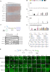
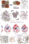
. 2025 Aug 22;11(34):eady8610.
doi: 10.1126/sciadv.ady8610. Epub 2025 Aug 22.

Characterization of the glycoproteins of fish and amphibian influenza B-like viruses

Affiliations

Characterization of the glycoproteins of fish and amphibian influenza B-like viruses

Gagandeep Singh et al. Sci Adv. .

Abstract

Influenza-like virus sequences previously identified in fish and amphibians cluster as a sister clade of influenza B viruses but remain largely uncharacterized. We demonstrate that salamander influenza-like virus (SILV) hemagglutinin (HA) is functionally divergent from influenza B virus HA and does not bind to α2,3- and α2,6-linked sialic acids. However, the HAs of Siamese algae-eater influenza-like virus (SAEILV) and chum salmon influenza-like virus (CSILV) bind to α2,3-linked sialic acid. Furthermore, SAEILV HA binds to sialyated Lewis X, is activated by human airway enzymes, and is fusogenic over a broad pH range. SAEILV neuraminidase (NA) has a highly conserved active site and a similar structure to other known NAs. We also determined the cryo-electron microscopy structure of the HA of a previously described virus from the same sister clade, the Wuhan spiny eel influenza virus (WSEIV). No cross-reactive antibodies against these HAs or NAs were found in human serum, suggesting that humans are immunologically naïve to these viruses.


Figures
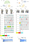
Fig. 1.
Fig. 1.. Phylogenetic and comparative sequence analysis of SILV, SAEILV, and CSILV glycoproteins.
Phylogenetic analysis of the SILV, SAEILV, and CSILV HA and NA amino acid sequences and representative sequences from influenza A and B virus HAs (A) and NAs (B) were performed by the maximum likelihood method. The scale bar shows estimated amino acid substitutions per site. Conserved amino acid residues of the SILV HA and NA (C and F), SAEILV HA and NA (D and G), CSILV HA and NA (E and H), respectively, relative to influenza B virus HA and NA are highlighted in blue on top of the publicly available structure of influenza B/Brisbane/60/2008 HA (PDB 4FQM) and NA (PDB 4CPL), visualized by ChimeraX. The HA RBS, central stem (CS) epitope, and NA active site are circled in red, orange, and purple. Amino acid sequence alignment of the SILV HA and NA, SAEILV HA and NA, CSILV HA and NA, and WSEIV HA and NA are displayed against influenza B/Malaysia/2506/2004 HA and NA. (I and J) Key features are shown in different colors. Asterisks indicate identical amino acids. SDS-PAGE analysis of recombinant expressed SILV, SAEILV, and CSILV HAs (K) and SAEILV NAs (L) and influenza B/Malaysia/2506/2004 HA and NA under nondeglycosylated and deglycosylated conditions.
Fig. 2.
Fig. 2.. Functional profile of SILV, SAEILV, and CSILV HAs.
(A) Hemagglutination assays were performed using recombinant HAs from SILV, SAEILV, and CSILV with turkey and chicken RBCs. HA proteins were added at 10 μg/ml and serially diluted twofold. (B to E) Glycan microarray analysis was used to assess binding of recombinant SILV, SAEILV, and CSILV HAs to synthetic glycans with α2,3- or α2,6-linked sialic acids (Neu5Ac), including linear, biantennary, triantennary, and SLex structures. B/Netherlands/2914/2015 HA was used as a positive control. Glycans A to M and O to W represent linear and biantennary structures, whereas glycans 1 to 22 represent triantennary N-glycans with LacNAc extensions. Glycans are color coded by terminal motif: yellow (no NeuAc), pink (α2,6-NeuAc), white (α2,3-NeuAc), blue (Lex), and black (SLex). Bars with dual colors indicate distinct epitopes on different arms. Data are shown as mean RFUs ± SD. (F) HA cleavage was assessed in HEK293T cells cotransfected with plasmids expressing full-length HAs and human airway proteases. After 48 hours, cleavage of HA0 to HA1 was evaluated by Western blot. Cells transfected with pCAGGS-HA, treated or untreated with TPCK-treated trypsin, served as controls. (G) Cell-cell fusion was assessed in HeLa cells expressing the respective HAs. Following trypsin treatment and low-pH exposure, polykaryon formation was evaluated. Representative images show fusion induced by SILV, SAEILV, CSILV, and B/Malaysia/2506/2004 HAs. White arrows indicate polykaryons, with insets showing magnified views of the fused cells. UT, untransfected.
Fig. 3.
Fig. 3.. WSEIV HA cryo-EM structure resembles influenza B virus HA.
(A) Cryo-EM map and model of WSEIV HA showing the HA1/HA2 subunits, fusion peptide, RBS pocket, and N-linked glycans. (B) Models of WSEIV HA and B/Brisbane/60/2008 HA (PDB 4FQM) are aligned by the central helix of HA2. (C) The surface of WSEIV HA and B/Brisbane/60/2008 HA is colored by electrostatic potential using ChimeraX. (D) Key residues within the RBS of WSEIV HA and B/Brisbane/60/2008 HA. Ab initio reconstruction maps and selected 2D classes of SAEILV HA (E) and SILV HA (F) from nsEM.
Fig. 4.
Fig. 4.. NA specific activities and cleavage specificities of the SAEILV NA.
(A) NA enzymatic activity of recombinant SAEILV and B/Malaysia/2506/2004 NA proteins was examined via ELLA using fetuin, at four different temperatures: 4°, 20°, 33°, and 37°C. The curves indicate absorbance measured at 490 nm with error bars indicating the SD. OD, optical density. (B) Specific enzymatic activity (inverse of half-maximum lectin binding) is shown for each individual NA at each temperature. (C) Inhibitory susceptibility to three NA inhibitors (oseltamivir, peramivir, and zanamivir) was evaluated in an ELLA-based NA inhibition assay. (D) Specific activity of SAEILV and B/Malaysia/2506/2004 NA as determined by ELLA using different glycoprotein-lectin combinations normalized to the fetuin-ECA.
Fig. 5.
Fig. 5.. SAEILV NA cryo-EM structure resembles influenza B virus NA.
(A) Cryo-EM map of SAEILV NA showing the tetrameric NA head, active site, and N-linked glycan. (B) Aligned models of SAILV NA, WSEIV NA (PDB 7XVU), and B/Brisbane/60/2008 NA (PDB 4CPL) showing conserved secondary structures, key active-site residues (B NA numbering), and underside glycans among three NAs. (C) The surface and the active-site pockets of SAEILV NA, WSEIV NA, and B/Brisbane/60/2008 NA are colored by electrostatic potential using ChimeraX. (D) Cryo-EM map and model showing the conserved calcium binding pocket in SAEILV NA. (E) Aligned models of SAEILV NA and 1G01 + A/California/04/2009 (H1N1) showing that 1G01 HCDR3 clashes with SAEILV NA Y239.
Fig. 6.
Fig. 6.. Binding profiles of the anti–influenza B virus HA and NA mAbs.
(A and C) Broadly cross-reactive anti–influenza B virus HA and NA human and mouse mAb binding to recombinant protein (SILV, SAEILV, CSILV, and B/Malaysia/2506/2004 HA and SAEILV and B/Malaysia/2506/2004 NA) in ELISA. (B) Anti–influenza B virus human and mouse mAb binding to cell surface expressed NAs of SILV, SAEILV, CSILV, and B/Malaysia/2506/2004 by flow cytometry. (D) Cross-reactive antibody profile of sera from postseasonal influenza vaccination recipients. Antibodies against SILV, SAEILV, CSILV HA, and SAEILV NA were determined via ELISA. Mopeia virus glycoprotein was used as the negative control for baseline establishment.

Update of

Similar articles

References

    1. Krammer F., Smith G. J. D., Fouchier R. A. M., Peiris M., Kedzierska K., Doherty P. C., Palese P., Shaw M. L., Treanor J., Webster R. G., García-Sastre A., Influenza. Nat. Rev. Dis. Primers. 4, 3 (2018). - PMC - PubMed
    1. D. M. Knipe, P. Howley, Fields Virology 6th Edition (Lippincott Williams & Wilkins, 2013).
    1. Short M. R. K. R., Verhagen J. H., van Riel D., Schrauwen E. J. A., van den Brand J. M. A., Mänz B., Bodewes R., Herfst S., One health, multiple challenges: The inter-species transmission of influenza A virus. One Health 1, 1–13 (2015). - PMC - PubMed
    1. Halwe N. J., Hamberger L., Sehl-Ewert J., Mache C., Schön J., Ulrich L., Calvelage S., Tönnies M., Fuchs J., Bandawane P., Loganathan M., Abbad A., Carreño J. M., Bermúdez-González M. C., Simon V., Kandeil A., El-Shesheny R., Ali M. A., Kayali G., Budt M., Hippenstiel S., Hocke A. C., Krammer F., Wolff T., Schwemmle M., Ciminski K., Hoffmann D., Beer M., Bat-borne H9N2 influenza virus evades MxA restriction and exhibits efficient replication and transmission in ferrets. Nat. Commun. 15, 3450 (2024). - PMC - PubMed
    1. Osterhaus A. D., Rimmelzwaan G. F., Martina B. E., Bestebroer T. M., Fouchier R. A., Influenza B virus in seals. Science 288, 1051–1053 (2000). - PubMed

MeSH terms

Substances

LinkOut - more resources