Review
. 2025 Aug;15(4):e70042.
doi: 10.1002/cph4.70042.

Crosstalk Between Microbiome and Ferroptosis in Diseases: From Mechanism to Therapy

Affiliations
Review

Crosstalk Between Microbiome and Ferroptosis in Diseases: From Mechanism to Therapy

Si-Qi Ding et al. Compr Physiol. 2025 Aug.

Abstract

The human microbiome is a unique organ and maintains host immunomodulation and nutrient metabolism. Structural and functional microbiome alterations are commonly known as dysbiosis, which is strongly associated with disease progression. Ferroptosis is a novel iron-dependent cell death mode characterized by intracellular iron accumulation, increased reactive oxygen species (ROS), and lipid peroxidation (LPO). Importantly, the complex crosstalk between the microbiome and ferroptosis in disease has attracted considerable research attention. The microbiome influences ferroptosis by regulating host iron homeostasis, mitochondrial metabolism, and LPO, among many other pathways. Thus, the in-depth analysis of microbiome-ferroptosis crosstalk and associated mechanisms could provide new strategies to treat human diseases. Therefore, understanding this crosstalk is critical. Here, we systematically explore the associations between gut microbiome and ferroptosis across multiple diseases. We show that the oral microbiome also influences disease progression by regulating ferroptosis. Furthermore, we provide a potential for certain disease therapies by targeting the crosstalk between the microbiome and ferroptosis.

Keywords: fecal microbiota transplantation; ferroptosis; gut‐organ‐axis; microbiome; probiotics.


Conflict of interest statement

The authors declare no conflicts of interest.

Figures

FIGURE 1
FIGURE 1
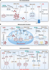
Ferroptosis driver or suppressor pathways. Ferroptosis driver pathways: TFR and DMT1 transport Fe3+ into cells and convert it to Fe2+. Intracellular iron is exported to extracellular sites by FPN. Fe2+ released by intracellular ferritinophagy promotes ROS production and lipid peroxidation via Fenton reactions. Most intracellular Fe2+ is used by the mitochondria. Electrons produce superoxide from electron transfer chain complexes I and III leak or NOXs transfer, which can be reduced to H2O2 by SOD2. H2O2 also reacts with unstable iron to produce abundant ROS (Fenton reactions). Additionally, free iron overload causes uncontrolled mitophagy, resulting in high free iron, ROS, and lipid peroxide levels entering the cytoplasm and exacerbating ferroptosis. PUFA is catalyzed by ACSL4 to produce PUFA‐CoA, which produces PUFA‐PL under LPCAT3. PUFA‐PL produces many ROS molecules through POR and ALOX actions, causing LPO and promoting ferroptosis. Ferroptosis suppressor pathways: Ferroptosis inhibition pathways include: Xc‐GSH‐GPX4, FSP1‐CoQH2, DHODH‐CoQH2, GCH1‐BH4, MBOAT1/2‐MUFA, and SC5D‐7‐DH axis systems. SLC3A2 and SLC7A11 exchange glutamate and cystine, while reducing cystine to cysteine to synthesize GSH and promote GPX4 production. ACSL4, acyl‐CoA synthetase long‐chain family member 4; ALOX, lipoxygenase; CoQH2, ubiquinol; DMT1, divalent metal transporter 1; FPN, ferroportin; FSP1, ferroptosis suppressor protein 1; GPX4, glutathione peroxidase 4; GSH, glutathione; LPCAT3, lysophosphatidylcholine acyltransferase 3; LPO, lipid peroxidation; NOXs, NADPH oxidases; POR, oxidoreductase cytochrome P450 reductase; PUFA, polyunsaturated fatty acid; PUFA‐PL, polyunsaturated fatty acid‐phospholipid; ROS, reactive oxygen species; SLC3A2, solute carrier family 3 member 2; SLC7A11, solute carrier family 7a member 11; SOD2, superoxide dismutase 2; STEAP3, six‐transmembrane epithelial antigen of prostate 3.
FIGURE 2
FIGURE 2
General microbiome properties and the microbiome‐gut‐organ axis. (A) The microbiome is distributed across different parts of the body, such as the digestive, respiratory, urogenital tract, and skin systems, and also tumor tissues. Microbiome composition includes bacteria, archaea, viruses, phages, fungi, and parasites, their structural elements, and various metabolites (e.g., LPS, SCFAs, and BAs). (B) The gut microbiome communicates with the liver, brain, oral system, and bone through immune, inflammatory, bloodstream, and other pathway systems to establish microbiome‐gut‐organ axes. BAs, bile acids; FXR, farnesoid X receptor; GLUT1, glucose transporter‐1; HDAC, histone deacetylase; LPS, lipopolysaccharide; NOX2, NADPH oxidases 2; SCFAs, short‐chain fatty acids; TLR4, toll‐like receptor‐4.
FIGURE 3
FIGURE 3
Microbial regulation of ferroptosis. (A) The microbiome inhibits FPN expression by inducing hepcidin or inhibiting HIF‐2α expression, and inducing intracellular iron overload. DCA promotes DMT1 expression via HIF‐2α, leading to increased labile iron levels. Microbial metabolites promote ferritin storage. Butyrate promotes HIF‐1α‐NCOA4‐mediated ferritinophagy, resulting in high Fe2+ levels entering the LIP and producing ROS, thereby inducing ferroptosis. F. nucleatum induces ferroptosis by increasing unstable iron levels in cells. (B) LPS induces NCOA4‐mediated ferritinophagy to activate SFXN1, and transfers excess Fe2+ to mitochondria, resulting in ferroptosis. F. nucleatum ‐induced iron overload may cause mitochondrial dysfunction. Excess iron accumulation in the mitochondria leads to the ROS, LPO, and mitophagy‐mediated aggravation of intracellular labile iron, which further induces ferroptosis. Additionally, propionate promotes mitochondrial damage, fission, and mitophagy. CAT also inhibits mitophagy and oxidative stress by inhibiting HIF‐1α expression to ultimately inhibit ferroptosis. Bomidin and butyrate increase GPX4 expression by upregulating the KEAP1/NRF2 pathway, which reduces mitochondrial damage and subsequent LPO. Tumoral butyrate increases intracellular ROS levels and mitochondrial metabolic damage by decreasing SOD2 expression. (C) Propionate elevates PUFA‐PL levels by increasing ACSL4 expression, while CagA reduces ACSL4 levels. CagA promotes PUFA‐ePLs synthesis, causing ferroptosis sensitivity in cells. P. aeruginosa promotes lipid oxidation by enhancing ALOX expression to induce ferroptosis. LPS‐induced NOX2 activation increases oxidative stress. Nevertheless, CagA or glutamine reduces NOX4 or NOX1 expression, respectively, both of which decrease ROS production and inhibit ferroptosis. AIEC colonization may decrease GPX4 expression, thus aggravating LPO and ferroptosis. F. nucleatum inhibits ferroptosis by elevating GPX4. Butyrate depletes intracellular GPX4 by inhibiting the ATF3/SLC7A11 axis to promote lipid peroxidation and ferroptosis. IDA increases FSP1‐CoQH2 levels and contributes to ferroptosis resistance. ACSL4, acyl‐CoA synthetase long‐chain family member 4; AIEC, adherent‐invasive E. coli ; ALOX, lipoxygenase; ATF3, transcription factor 3; CagA, cytotoxin‐associated gene A; CAT, Capsiate; CoQH2, ubiquinol; DCA, deoxycholic acid; DMT1, divalent metal transporter 1; FPN, ferroportin; FSP1, ferroptosis suppressor protein 1; GPX4, glutathione peroxidase 4; HIF‐1α, hypoxia‐inducible factor‐1α; HIF‐2α, hypoxia‐inducible factor‐2α; IDA, trans‐3‐indoleacrylic acid; KEAP1, Kelch‐associated protein 1; LIP, labile iron pool; LPO, lipid peroxidation; LPS, lipopolysaccharide; NCOA4, nuclear receptor co‐activator 4; NOX1/2/4, NADPH oxidases 1/2/4; NRF2, nuclear factor erythroid 2‐related factor 2; P. aeruginosa , Pseudomonas aeruginosa ; PUFA‐ePLs, polyunsaturated ether phospholipids; PUFA‐PL, polyunsaturated fatty acid‐phospholipid; SFXN1, siderofexin; SOD2, superoxide dismutase 2.
FIGURE 4
FIGURE 4
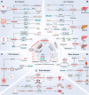
Crosstalk between the microbiome and ferroptosis in disease. The microbiome regulates ferroptosis in several ways to influence gut, liver, brain, oral, bone, and other diseases. ABL, alveolar bone loss; ACSL4, acyl‐CoA synthetase long‐chain family member 4; AD, Alzheimer's disease; AHR, aryl hydrocarbon receptor; AIEC, adherent‐invasive E. coli ; ALDH1A3, aldehyde dehydrogenase 1 family member A3; ALK5, activin receptor‐like kinase 5; ALOX, lipoxygenase; BAA, bromoacetic acid; BAs, bile acids; CAT, capsiate; CDCA, chenodeoxycholic acid; CRC, colorectal cancer; CXCL1, C‐X‐C motif chemokine ligand‐1; DAMPs, danger‐associated molecular patterns; DCA, deoxycholic acid; DMT1, divalent metal transporter 1; EPS, exopolysaccharides; FSP1, ferroptosis suppressor protein 1; GCDCA, glycochenodeoxycholate; GPX4, glutathione peroxidase 4; HCC, hepatocellular carcinoma; HIF‐1α, hypoxia‐inducible factor‐1α; HIF‐2α, hypoxia‐inducible factor‐2α; IBD, inflammatory bowel disease; ICC, intrahepatic cholangiocarcinoma; IDA, trans‐3‐indoleacrylic acid; IS, ischemic stroke; KEAP1, Kelch‐associated protein 1; LPC, lysophosphatidylcholine; LPS, lipopolysaccharide; MAFLD, metabolic dysfunction‐associated fatty liver disease; n‐3 PUFA, omega‐3 polyunsaturated fatty acids; NCOA4, nuclear receptor co‐activator 4; NOX1, NADPH oxidases 1; NRF2, nuclear factor erythroid 2‐related factor 2; NRF2, nuclear factor erythroid 2‐related factor 2; OA, oleanolic acid; OCA, obeticholic acid; OS, osteosarcoma; P. gingivalis , Porphyromonas gingivalis ; PD, Parkinson's disease; PPAR, peroxisome proliferator‐activated receptor; SCFAs, short‐chain fatty acids; SLC2A1, solute carrier family 2 member 1; TRPV1, transient receptor potential cation channel subfamily V member 1; UroC, urolithin C.

Similar articles

References

    1. Abed, J. , Maalouf N., Manson A. L., et al. 2020. “Colon Cancer‐Associated <styled-content style="fixed-case"> Fusobacterium nucleatum </styled-content> May Originate From the Oral Cavity and Reach Colon Tumors via the Circulatory System.” Frontiers in Cellular and Infection Microbiology 10: 400. - PMC - PubMed
    1. Aggarwal, N. , Kitano S., Puah G. R. Y., Kittelmann S., Hwang I. Y., and Chang M. W.. 2023. “Microbiome and Human Health: Current Understanding, Engineering, and Enabling Technologies.” Chemical Reviews 123, no. 1: 31–72. - PMC - PubMed
    1. Ahlqvist, K. J. , Leoncini S., Pecorelli A., et al. 2015. “MtDNA Mutagenesis Impairs Elimination of Mitochondria During Erythroid Maturation Leading to Enhanced Erythrocyte Destruction.” Nature Communications 6: 6494. - PubMed
    1. Ahuja, S. , and Lazar I. M.. 2021. “Systems‐Level Proteomics Evaluation of Microglia Response to Tumor‐Supportive Anti‐Inflammatory Cytokines.” Frontiers in Immunology 12: 646043. - PMC - PubMed
    1. Albuquerque‐Souza, E. , and Sahingur S. E.. 2022. “Periodontitis, Chronic Liver Diseases, and the Emerging Oral‐Gut‐Liver Axis.” Periodontology 2000 89, no. 1: 125–141. - PMC - PubMed

LinkOut - more resources