. 2025 Aug 22;16(1):7828.
doi: 10.1038/s41467-025-63056-3.

Anti-restriction functions of injected phage proteins revealed by peeling back layers of bacterial immunity

Affiliations

Anti-restriction functions of injected phage proteins revealed by peeling back layers of bacterial immunity

Sukrit Silas et al. Nat Commun. .

Abstract

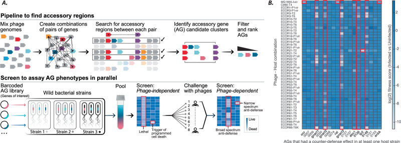
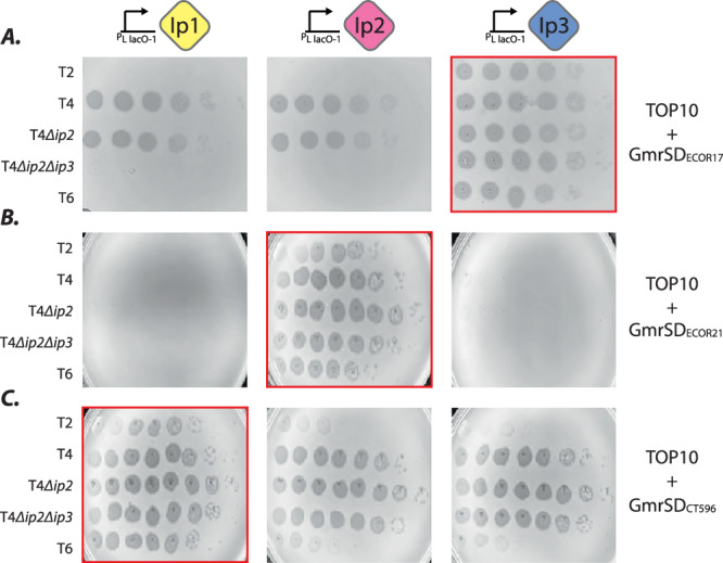
Virus-host competition drives evolution of diverse antivirus defenses, but how they co-operate in wild bacteria and how bacteriophages circumvent host immunity remains poorly understood. Here, using a functional screening platform to systematically explore the functions of phage accessory genes, we describe how cell-surface barriers can obscure the phenotypes of intracellular defenses in E. coli isolates. LPS modification emerged as a major theme, with the discovery of several small phage proteins that modify specific O-antigen structures, removing barriers to phage adsorption. Bypassing O-antigen in wild E. coli strains revealed another layer of defense: Type IV restriction endonucleases (RE) that target modified DNA of T-even phages (T2, T4, T6). We further show how injected proteins Ip2 and Ip3 of T4 inhibit distinct subtypes of these Type IV REs. Extensive variability in Type IV REs likely drives the emergence of subtype-specific inhibitors through multiple rounds of adaptation and counter-adaptation.


Conflict of interest statement

Competing interests: S.S. is co-founder and equity holder in BillionToOne Inc., BK Labs Inc., and CK Labs Inc., and a scientific advisory board member of Junevity Inc. J.B.D. is a scientific advisory board member of SNIPR Biome, Excision Biotherapeutics, and LeapFrog Bio, consults for BiomX, and is a scientific advisory board member and co-founder of Acrigen Biosciences and ePhective Therapeutics. The Bondy-Denomy lab received research support from Felix Biotechnology.

Figures

Fig. 1
Fig. 1. High-throughput screening to find anti-defense phage AGs.
A Schematic of a computational pipeline to identify phage AGs (described in detail elsewhere), and high-throughput screening platform to assay AG functions in multiple wild strains of bacteria. B Heatmap of log2-transformed fitness scores comparing infected and uninfected samples. Of 196 AGs tested, only AGs with a host-sensitizing phenotype are shown on the X-axis. All genes listed on the X-axis were tested in at least one host by plaque assays performed in parallel by two different experimenters, and hits thus validated were further verified by additional plaque assays in triplicate. Red boxes/bold names highlight counter-defense AGs selected for further study. Named AGs are orf1:ocr, orf48:gnarl1, orf63:gnarl2, orf87:ral, orf92:gnarl3, orf116:abc1, orf143:ip2 and orf169:ardA. Mean fitness scores are presented from three independent replicates of the entire screen (see Supplementary Data 1).
Fig. 2
Fig. 2. Phage AGs that compromise O-antigen barrier defenses on the cell surface.
A Schematic of follow-up transposon screens, performed in native hosts without AGs. Graphs show fitness of transposon mutants in infected (Y-axis) and uninfected (X-axis) Tn5-libraries from three representative hosts challenged by T5. Red dots are gene-disruptions that lower bacterial fitness upon infection (e.g., disruptions in O-antigen biosynthesis). B Median T5 infection scores in wild hosts ECOR1 (green), ECOR3 (orange), ECOR22 (blue) with gnarl genes (orf48:gnarl1, orf63:gnarl2, orf92:gnarl3) repressed (left) or induced (right). Infection scores from triplicate plaque assays are depicted by circles. C Lipopolysaccharide (LPS) visualized by electrophoresis (n = 1) from host-AG combinations in (B) where AGs produced a phage-sensitizing effect. (D) As in (B), Median T5 infection scores with or without gnarl3 in ECOR hosts, with UDP-glucose biosynthesis pathway genes galU, galE, galF, and Enterobacterial Common Antigen (ECA) precursor wecB cloned onto a plasmid and overexpressed. E GST-immunoprecipitation and western blotting (n = 2) with anti-GST (white background) and anti-FLAG (black background) antibodies to test association of Gnarl3 and GalU proteins. GalU (lane 2) or GST-GalU (lanes 1, 3) expressed from a plasmid (pBAD), and FLAG-Gnarl3 (lanes 1, 2) expressed from a single-copy chromosomal insertion (pLlacO-1). Input lysate controls were loaded onto the same gels (lanes 1-3) as the GST-IP eluates (lanes 4-6). Anti-FLAG images are from the same Western Blot, but eluate lanes (4-6) were visualized separately with longer exposure times as FLAG signal in input lysates (1-3) was substantially brighter. Source data for (C, E) are provided as a Source Data file.
Fig. 3
Fig. 3. A Type IV RE embedded in a BREX locus.
A Fitness of transposon-mutants in infected and uninfected Tn5-libraries of ECOR21 challenged by various phages. B Schematic of ECOR21 defense against T2 through both barrier defenses and a defense system which is putatively blocked by orf143/Ip2. C LPS visualized (n = 1) from WT ECOR21 (left), and ECOR21 with the O-antigen biosynthesis initiator wecA deleted by allelic exchange (right). D Whole-genome transposon-mutagenesis screens as in (A) but with ECOR21::∆wecA. E Variants of the BREX/GmrSD operon cloned onto a plasmid along with their native promoters and tested in a lab strain of E. coli (TOP10) against T-even phages (T2, T4, T6) in triplicate plaque assays. Removal of the PglX gene inactivates BREX. Active-site mutations that disable GmrSD are shown as two parallel red bars (GmrSD* double mutant; D474A, H475A). Ocr, Ip1 and Ip2 are expressed from single-copy chromosomal insertions. Bar graphs show median log10 infection scores. F GmrSD and its inactive variant GmrSD* cloned onto a plasmid and tested in TOP10 for restriction activity against T-even phages as in (E). “pBAD” indicates the defense construct was overexpressed from the plasmid (no “pBAD”: same promoter but no arabinose induction). Infection scores from triplicate plaque assays are depicted by circles. Source data for (C) are provided as a Source Data file.
Fig. 4
Fig. 4. A Type IV RE with a GmrS/GmrSD doublet.
A Fitness of the same transposon-mutant library of ECOR17 (2.31e7 cfu) challenged by various amounts of a ~ 5.5e9 pfu/ml lysate of phage T4∆ip2ip3. Tn5 insertions that compromise phage resistance (red dots) in low-phage-input screens are listed in Supplementary Data 3. B Schematic of ECOR17 locus that encodes defense system putatively blocked by Ip3. C Plaque assays to measure anti-phage activity of GmrS1 and GmrSD2 components individually (with overexpression) and of the full system (without overexpression). T4 and T4∆ip2 phages naturally encode Ip3. D Plaque assays to measure anti-phage activity of constructs (without overexpression) with truncated GmrS1 or GmrSD2 with E582A H583A mutations in its active site. Images in (C), (D) are representative images from plaque assays performed in triplicate.
Fig. 5
Fig. 5. Injected proteins (ip) from T4 phage block specific GmrSD variants.
Plaque assays to measure anti-phage activity of (A) GmrSDECOR17 (CLS_003/004) (B) GmrSDECOR21 (CLS_010), and (C) GmrSDCT596 (CLS_006) with heterologous expression of Ip1-3 (red borders indicate GmrSD variant is paired with its ip inhibitor). ip genes are expressed from single-copy chromosomal insertions. Ip1 is naturally present in T4, T4∆ip2, and T4∆ip2ip3, Ip2 in T4, and Ip3 in T4 and T4∆ip2. All plasmid-encoded GmrSD genes were tested without over-expression.

Similar articles

References

    1. Doron, S. et al. Systematic discovery of antiphage defense systems in the microbial pangenome. Science359, eaar4120 (2018). - PMC - PubMed
    1. Gao, L. et al. Diverse enzymatic activities mediate antiviral immunity in prokaryotes. Science369, 1077–1084 (2020). - PMC - PubMed
    1. Millman, A. et al. An expanded arsenal of immune systems that protect bacteria from phages. Cell host microbe30, 1556–1569 e1555 (2022). - PubMed
    1. Vassallo, C. N., Doering, C. R., Littlehale, M. L., Teodoro, G. I. C. & Laub, M. T. A functional selection reveals previously undetected anti-phage defence systems in the E. coli pangenome. Nat. Microbiol7, 1568–1579 (2022). - PMC - PubMed
    1. Rousset, F. et al. Phages and their satellites encode hotspots of antiviral systems. Cell host microbe30, 740–753.e745 (2022). - PMC - PubMed

MeSH terms

LinkOut - more resources