

Microglial activation is inhibited by selective anti-seizure medications
- PMID: 40848130
- PMCID: PMC12374903
- DOI: 10.1007/s00011-025-02076-7
Microglial activation is inhibited by selective anti-seizure medications
Abstract
Objective: To investigate the anti-inflammatory properties of anti-seizure medications (ASMs) administered to patients with drug-resistant epilepsy (DRE) and the role of sodium channels in microglial activation.
Material: Primary microglia monocultures from mice brains.
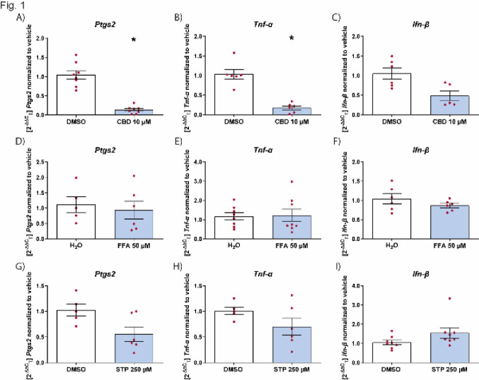
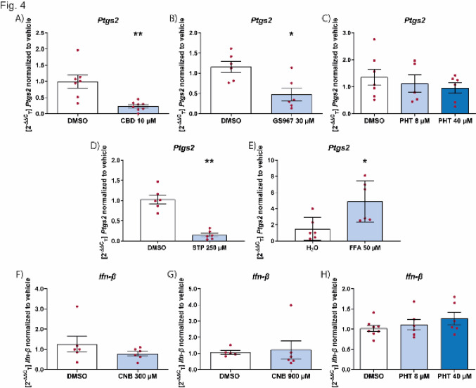
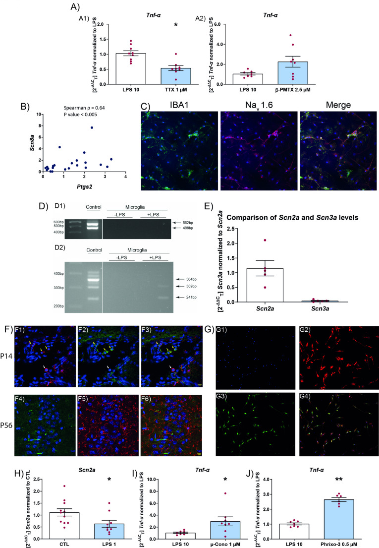
Treatment: Microglia were activated with 10 μg/mL lipopolysaccharide (LPS) or polyinosinic:polycytidylic acid (poly I:C) and pre- (45 min ASM then 2 h ASM plus stimulus) or post- (2 h stimulus then 24 h only ASM) treated with ASMs. Microglia were treated with cannabidiol (10 μM), stiripentol (250 μM), fenfluramine (50 μM), phenytoin (8 and 40 μM), cenobamate (300 and 900 μM), or the small molecule sodium channel blocker GS967 (10 and 30 μM). The sodium channel modulators tetrodotoxin (1 μM), µ-conotoxin KIIIA (1 μM), and β-pompilidotoxin (0.5 μM) were also applied.
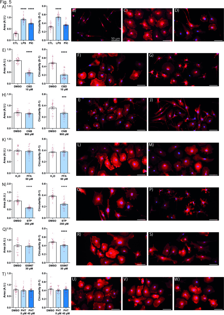
Methods: Microglia activation was quantified through measurements of Ptgs2 (Cox2), Tnf-α, and Ifn-β induction by RT-qPCR and of cell morphology by immunocytochemistry. Expression of sodium channels in microglia was studied using PCR, RT-qPCR, immunohisto- and immunocytochemistry. Mann Whitney test and the Kruskal-Wallis test with Dunn's multiple comparisons post-test were used.
Results: ASMs have a differential effect on microglial activation. Uniquely, cenobamate inhibited the induction of Ifn-β and made the cells less amoeboid. The voltage gated sodium channel Nav1.2 is expressed by microglial cells and its expression levels change with microglial inflammatory response. Toxins that block sodium channels modulated microglial activation.
Conclusions: ASMs, applied to patients with DRE, have a differential ability to reduce microglial activation and pro-inflammatory microglial morphology in vitro. Moreover, sodium channel blockage modulates inflammation through microglia activation. Taken together these results suggest, that further investigation of patient's immune response to ASMs could be important.
Keywords: Anti-seizure medication; Epilepsy; Inflammation; Microglia.
© 2025. The Author(s).
Conflict of interest statement
Declarations. Conflict of interest: Angela M. Kaindl has served as a consultant for Angelini Pharma, Desitin, Jazz Pharmaceuticals, and UCB. The remaining authors have no conflicts of interest. Consent for patients: Not applicable. Ethical approval: We confirm that we have read the Journal’s position on issues involved in ethical publication and affirm that this report is consistent with those guidelines. All animal experiments were carried out in accordance to the national ethic principles (registration no. T0344/12, State Office for Health (LAGeSo), Berlin, Germany).
Figures


Similar articles
-
Nimodipine reduces microglial activation in vitro as evidenced by morphological phenotype, phagocytic activity and high-throughput RNA sequencing.Br J Pharmacol. 2025 Aug;182(16):3800-3817. doi: 10.1111/bph.70060. Epub 2025 Apr 21. Br J Pharmacol. 2025. PMID: 40258609
-
Prescription of Controlled Substances: Benefits and Risks.2025 Jul 6. In: StatPearls [Internet]. Treasure Island (FL): StatPearls Publishing; 2025 Jan–. 2025 Jul 6. In: StatPearls [Internet]. Treasure Island (FL): StatPearls Publishing; 2025 Jan–. PMID: 30726003 Free Books & Documents.
-
Microglial priming by IFN-γ involves STAT1-mediated activation of the NLRP3 inflammasome.CNS Neurosci Ther. 2024 Oct;30(10):e70061. doi: 10.1111/cns.70061. CNS Neurosci Ther. 2024. PMID: 39392762 Free PMC article.
-
Monotherapy treatment of epilepsy in pregnancy: congenital malformation outcomes in the child.Cochrane Database Syst Rev. 2023 Aug 29;8(8):CD010224. doi: 10.1002/14651858.CD010224.pub3. Cochrane Database Syst Rev. 2023. PMID: 37647086 Free PMC article.
-
Cenobamate add-on therapy for drug-resistant focal epilepsy.Cochrane Database Syst Rev. 2024 Aug 1;8(8):CD014941. doi: 10.1002/14651858.CD014941.pub2. Cochrane Database Syst Rev. 2024. PMID: 39087564 Free PMC article.
References
-
- Brodie MJ, Shorvon SD, Canger R, Halász P, Johannessen S, Thompson P, et al. Commission on European Affairs: appropriate standards of epilepsy care across Europe. ILEA Epilepsia. 1997;38:1245–50. - PubMed
-
- Victor TR, Hage Z, Tsirka SE. Prophylactic administration of cannabidiol reduces microglial inflammatory response to kainate-induced seizures and neurogenesis. Neuroscience. 2022;500:1–11. - PubMed
-
- Sills GJ, Rogawski MA. Mechanisms of action of currently used antiseizure drugs. Neuropharmacology. 2020;168:107966. - PubMed
-
- Villasana-Salazar B, Vezzani A. Neuroinflammation microenvironment sharpens seizure circuit. Neurobiol Dis. 2023;178:106027. - PubMed
MeSH terms
Substances
LinkOut - more resources
Full Text Sources
Research Materials
