. 2025 Aug 25;8(1):544.
doi: 10.1038/s41746-025-01948-w.

Culture-free detection of bacteria from blood for rapid sepsis diagnosis

Affiliations

Culture-free detection of bacteria from blood for rapid sepsis diagnosis

M Henar Marino Miguélez et al. NPJ Digit Med. .

Abstract

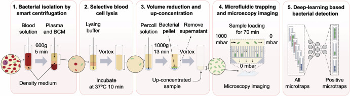
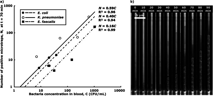
Approximately 50 million people suffer from sepsis yearly, and 13 million die from it. For every hour a patient with septic shock is untreated, their survival rate decreases by 8%. Therefore, rapid detection and antibiotic susceptibility profiling of bacterial agents in the blood of sepsis patients are crucial for determining appropriate treatment. Here, we introduce a method to isolate bacteria from whole blood with high separation efficiency through Smart centrifugation, followed by microfluidic trapping and subsequent detection using deep learning applied to microscopy images. We detected, within 2 h, E. coli, K. pneumoniae, or E. faecalis from spiked samples of healthy human donor blood at clinically relevant concentrations as low as 9, 7 and 32 colony-forming units per ml of blood, respectively. However, the detection of S. aureus remains a challenge. This rapid isolation and detection represents a significant advancement towards culture-free detection of bloodstream infections.


Conflict of interest statement

Competing interests: J.E. and W.W. have commercial interests in the diagnostic field. M.M., M.O., J.E., and W.W. have filed a patent application based on the method presented in this paper.

Figures

Fig. 1
Fig. 1. Workflow for bacterial detection from blood samples in five assay steps.
1 Isolation using smart centrifugation; 2 selective blood cell lysis; 3 volume reduction; 4 microfluidic trapping and microscopy imaging; and 5 deep-learning based bacterial detection. BCM is blood culture medium. Created in BioRender. Lab, E. (2025) https://BioRender.com/gyvhthfand in open-source software Inkscape.
Fig. 2
Fig. 2. Bacterial isolation from blood by smart centrifugation.
a Illustration of liquid and cell movement. The left and right tubes illustrate the positions of sample liquid, density medium, red blood cells and bacteria before and after smart centrifugation. The middle graph qualitatively illustrates the trajectories (solid lines) of bacteria (green) and red blood cells (red) during centrifugation, from a mixed state (left brackets) to a separated state (right brackets). The slopes of the lines are the particle sedimentation speeds, vRBC and vbac, and liquid interface, vint, and sedimentation interface velocity, vsed, derived in SI. b Blood cell removal efficiency, meaning the fraction of blood cells removed from the supernatant after centrifugation relative to the initial number of blood cells in the sample (n=3). c Bacterial isolation efficiency, meaning the number of colony-forming units in the supernatant after centrifugation relative to the initial number of colony-forming units in the spiked sample. Bar heights are mean; error bars are sd; n.s. and *** indicate significance levels p > 0.05 and p ≤ 0.001, respectively; the bacterial concentration C refers to the CFU /ml in the blood sample. Each data point corresponds to an individual experiment. A total of n = 70 samples were tested. The figures were created and edited using the open-source software Inkscape (a) and MATLAB R2021 (b) and (c).
Fig. 3
Fig. 3. Bacterial microtrapping.
a Overall assay performance, where dots represent the number of positive microtraps detected, N, for various bacterial concentrations in the 2.25 ml blood sample, C, and lines depict the least square fitting linear calibration curves N = ηC. Each point corresponds to an individual experiment. A total of n = 12 samples were tested. b Timelapse images of bacterial capture and growth in a single microtrap, showing the capture of one bacterium of K. pneumoniae 40 min after sample addition, followed by bacterial cell division. The figure was created with Microsoft Excel and edited and assembled using the open-source software Inkscape.
Fig. 4
Fig. 4. Deep learning model performance.
a Evaluating networks using downsampled data, testing on an increasing number of frames (each corresponding to 10 min). The heatmap shows the F1-Score of DinoV2. b Precision, recall, AUC (Area under the curve), and F1-Score of all models over time at full resolution, corresponding to the red dashed rectangle in part a Lines show mean, and shaded areas show standard deviations among the 30 retrainings. c Confusion matrix, performance metrics, and ROC (Receiver Operating Characteristics) curve of the DinoV2 model with the median AUC score among the 30 retrainings, at full resolution, evaluated at the final time (70 min). The figure was created using the open-source software Inkscape and the Python plotting library Matplotlib.

References

    1. Singer, M. et al. The third international consensus definitions for sepsis and septic shock (sepsis-3). J. Am. Med. Assoc.315, 801–810 (2016). - PMC - PubMed
    1. Rudd, K. E. et al. Global, regional, and national sepsis incidence and mortality, 1990-2017: analysis for the Global Burden of Disease Study. Lancet395, 200–211 (2020). - PMC - PubMed
    1. Thompson, K., Venkatesh, B. & Finfer, S. Sepsis and septic shock: current approaches to management. Intern. Med. J.49, 160–170 (2019). - PubMed
    1. Kumar, A. et al. Duration of hypotension before initiation of effective antimicrobial therapy is the critical determinant of survival in human septic shock. Crit. Care Med.34, 1589–1596 (2006). - PubMed
    1. Huerta, L. E. & Rice, T. W. Pathologic difference between sepsis and bloodstream infections. J. Appl. Lab. Med.3, 654–663 (2019). - PubMed

LinkOut - more resources