

Molecular imaging techniques in patients with persistent spinal pain syndrome type 2 - a systematic review and meta-analysis
- PMID: 40851058
- PMCID: PMC12375521
- DOI: 10.1186/s41824-025-00266-4
Molecular imaging techniques in patients with persistent spinal pain syndrome type 2 - a systematic review and meta-analysis
Abstract
Background: Persistent postoperative low back pain, including persistent spinal pain syndrome type 2 (PSPS-T2), is a global healthcare challenge that lacks objective phenotypic diagnostic criteria or validated biomarkers. Molecular imaging techniques (nuclear medicine) could aid in establishing more objective phenotypical parameters as they are able to visualize biological processes underlying several diseases even before anatomical changes are present. This review aims to provide an overview of the status quo of molecular imaging for the diagnosis of PSPS-T2.
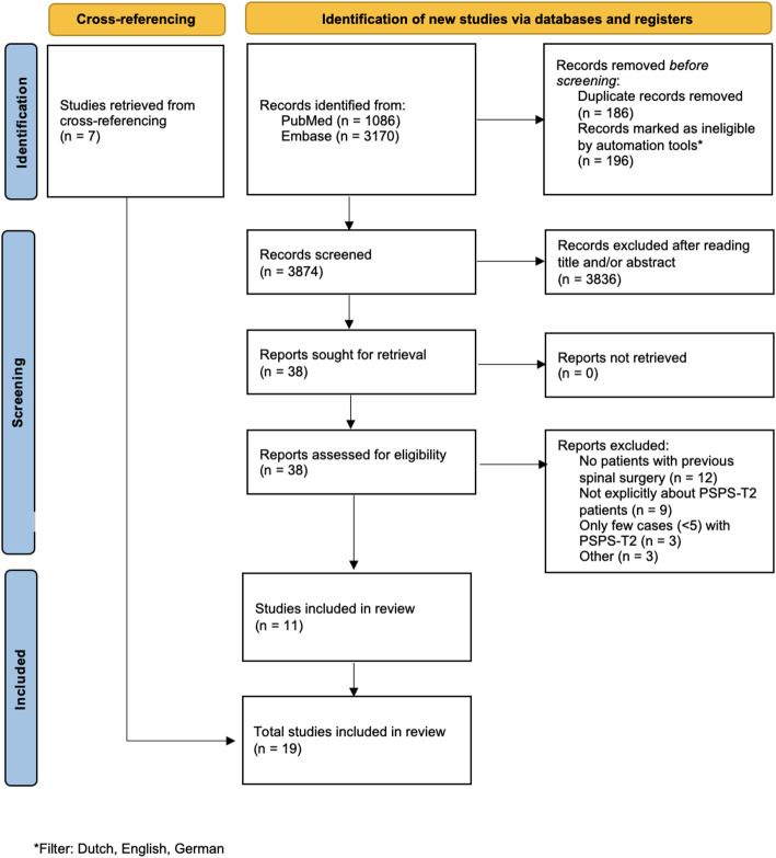
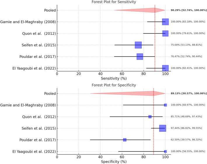
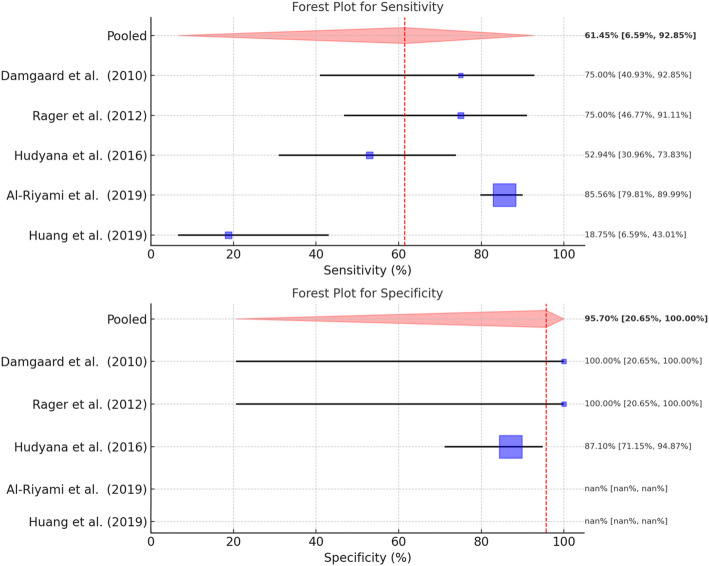
Method and results: An extensive search of PubMed and Embase was conducted to identify relevant studies that comprise imaging techniques using a radiopharmaceutical substance. Evidence reveals that these techniques can provide valuable insights into the underlying pathologies and mechanisms of PSPS-T2 by detecting pain generators that would have otherwise gone unnoticed. Moreover, the meta-analysis showed a pooled sensitivity of 90.3% (95% CI: 53–100%) and a specificity of 89.1% (95% CI: 31–100%) for [18F]NaF PET-CT, and a pooled sensitivity of 61.5% (95% CI: 7–93%) and specificity of 96% (95% CI: 21–100%) for diphosphonates SPECT-CT.
Conclusion: These findings suggest a potential utility in identifying those who are likely to benefit from surgical re-intervention. This illustrates the potential of molecular imaging in establishing a personalized-medicine approach. However, the retrospective design and the limited sample sizes are among the limitations of the included studies and further research is needed to unravel the potential of molecular imaging techniques as a tool to detect phenotypical biomarkers and to optimize patient care in patients with persistent postoperative low back pain, including PSPS-T2.
Keywords: Back pain; Chronic pain; Molecular imaging; Nuclear imaging; Persistent spinal pain syndrome type 2; post-surgical pain.
Conflict of interest statement
Declarations. Ethical approval and consent to participate: Not applicable. Consent for publication: Not applicable. Competing interests: The authors declare that they have no competing interests.
Figures
Similar articles
-
Percutaneous and Endoscopic Adhesiolysis in Managing Low Back and Lower Extremity Pain: A Systematic Review and Meta-analysis.Pain Physician. 2016 Feb;19(2):E245-82. Pain Physician. 2016. PMID: 26815254
-
Effectiveness of spinal endoscopic adhesiolysis in post lumbar surgery syndrome: a systematic review.Pain Physician. 2009 Mar-Apr;12(2):419-35. Pain Physician. 2009. PMID: 19305488
-
The Predicted Outcome of Spinal Cord Stimulation in Patients With a Psychopathological Disorder and Persistent Spinal Pain Syndrome Type 2: A Systematic Review From 2009 to 2021.Neuromodulation. 2024 Jan;27(1):59-69. doi: 10.1016/j.neurom.2023.11.004. Epub 2023 Dec 19. Neuromodulation. 2024. PMID: 38127048
-
Spinal cord stimulation for chronic back and leg pain and failed back surgery syndrome: a systematic review and analysis of prognostic factors.Spine (Phila Pa 1976). 2005 Jan 1;30(1):152-60. doi: 10.1097/01.brs.0000149199.68381.fe. Spine (Phila Pa 1976). 2005. PMID: 15626996
-
Efficacy of Epidural Injections in Managing Chronic Spinal Pain: A Best Evidence Synthesis.Pain Physician. 2015 Nov;18(6):E939-1004. Pain Physician. 2015. PMID: 26606031
References
-
- Acosta FL, Shapovalov VS, Lobo BM, Liker MA (2021) Radiographic evaluation of low back pain after lumbar fusion using Technetium-99 single-photon emission computed tomography: impact on clinical decision-making and outcome. Interdisciplinary Neurosurg 24:101089
-
- Adams C, Banks KP (2022) Bone scan. StatPearls. Treasure Island (FL): StatPearls publishing copyright © 2022. StatPearls Publishing LLC.
-
- Al-Riyami K, Vöö S, Gnanasegaran G, Pressney I, Meir A, Casey A et al (2019) The role of bone SPECT/CT in patients with persistent or recurrent lumbar pain following lumbar spine stabilization surgery. Eur J Nucl Med Mol Imaging 46(4):989–998 - PubMed
-
- Berquist TH (2006) Imaging of the postoperative spine. Radiol Clin North Am 44(3):407–418 - PubMed
Publication types
LinkOut - more resources
Full Text Sources