. 2025 Jul-Aug;73(7-8):289-314.
doi: 10.1369/00221554251365996. Epub 2025 Aug 25.

Expression Diversity in Endocrine Cells and Between Species Revealed by Novel Synthetic Peptide Antibodies Recognizing the Neuroendocrine Protein 7B2

Affiliations

Expression Diversity in Endocrine Cells and Between Species Revealed by Novel Synthetic Peptide Antibodies Recognizing the Neuroendocrine Protein 7B2

Shota Kikuchi et al. J Histochem Cytochem. 2025 Jul-Aug.

Abstract

The neuroendocrine protein 7B2 plays a crucial role in the maturation and activity regulation of prohormone convertase 2 (PC2). To elucidate the relationship between 7B2 and PC2 expression in endocrine tissues, we generated synthetic peptide antibodies in guinea pigs. The antigenic peptide sequences were selected to correspond to three different positions in the rat amino acid (aa) sequence: The N-terminal aa 1-14 is situated immediately following the signal sequence, the middle aa 77-90 contains a part of the proPC2 activation domain, and the C-terminal aa 156-168 functions to suppress PC2 activity. These antibodies demonstrated specific reactivity across a diverse array of animal species. The reactivity of these antibodies differed, suggesting that the molecular form of 7B2 differs depending on the endocrine cell, and a different expression pattern was demonstrated in rat and dog pituitary intermediate cells. The colocalization of 7B2 and PC2 in prolactin (PRL) granules in rat pituitary mammotrophs supports the interaction between these proteins. However, the expression intensities of these proteins did not correspond, and epitope-related disparities were detected. These results may be indicative of alterations in the molecular state associated with the dynamics of the interaction between 7B2 and PC2.

Keywords: adrenal medulla; gastrointestinal endocrine cell; pancreatic islets; peptide hormone; pituitary intermediate lobe; processing enzyme; secretogranin V; thyroid gland.


Conflict of interest statement

Competing InterestsThe author(s) declared no potential conflicts of interest with respect to the research, authorship, and/or publication of this article.

Figures

Figure 1.
Figure 1.
Amino acid (aa) sequences of 7B2 in seven species, and the locations of the antigenic peptides and functional domains. The positions of the synthetic peptide antigens are indicated as aa 1–14 (yellow), aa 77–90 (pink), and aa 156–168 (blue). The antigenic sites aa 23–39 (green) of the commercially available rabbit antibody, ab22699, are also shown. Human aa H120–P131 (H121–P132, as indicated by the lower dotted line) has been demonstrated to be involved in the facilitative activity of proPC2. Specifically, Y130 ($) has been identified as a critical component for 7B2 to function as an auxiliary protein for PC2. The T73 and T111 phosphorylation sites (#) involved in the interaction with proPC2 are shown. The domain Rat aa G86–Q122 (indicated by the lower wavy line) has been identified as a domain necessary for the activation of proPC2., The region subsequent to the furin cleavage site (RKRR, indicated by the red arrowhead) is the CT-peptide, which has been shown to inhibit the activation of proPC2. Two potential cleavage sites (KR and KK) of PC2 in the CT-peptide are indicated by black arrowheads. These cleavage sites have been identified as playing a critical role in the inactivation of the CT-peptide. The amino acids that did not correspond to the seven species are indicated by gray characters.
Figure 2.
Figure 2.
Immunoblot analysis of endocrine tissues. Tissue protein extracts (20 μg) were utilized for SDS–PAGE, followed by immunoblotting using rabbit antibody (ab22699) and three guinea pig #1–14, #77–90, and #156–168 antibodies. In the bovine adrenal medulla, #77–90 demonstrated high reactivity; conversely, low reactivity was observed in rat and dog whole adrenal extracts. The lane control encompasses PC2 and α-tubulin reactivities. Abbreviations: kDa, kilodalton.
Figure 3.
Figure 3.
PC2 and 7B2 expression in rat and canine pituitary glands. (A and K) HE staining of the rat and dog pituitary glands. (B and L) PC2 demonstrates notable reactivity in the intermediate part and scattered positive cells in the distal part of the adenohypophysis. (C and M) 7B2 labeled with ab22699 demonstrated a positive reaction in the adenohypophysis and neurohypophysis. However, the immunoreactive pattern exhibited a discrepancy from that of PC2, and a species-related difference was detected in the intermediate section. In the #1–14 labeling experiment, the number of positive cells in the adenohypophysis minimal was low in rats (E), yet a congruent pattern to ab22699 labeling was observed in dogs (O). (G and Q) In #77–90 labeling, a congruent pattern to that of ab22699 labeling was observed in both rats and dogs. (I and S) In the #156–168 labeling experiment, a negligible reaction was identified in the distal part of the rat and canine adenohypophysis. In contrast, reactions were observed in the canine intermediate part, but not in the rat intermediate part. Negative controls were reacted with normal rabbit IgG (Rb-IgG) (D and N) or pre-absorbed antibodies with immunogen peptides, aa 1–14 (F and P), aa 77–90 (H and R), and aa 156–168 (J and T). Abbreviations: Dis, distal part of adenohypophysis; Int, intermediate part of adenohypophysis; Neu, neurohypophysis. Scales, 100 µm. (U and V) The figure shows the percentage of 7B2 immunoreactive areas in the pituitary distal and intermediate parts of rats (blue bars) and dogs (orange bars). Data are presented as averages, with values from four non-overlapping images indicated by dots. *p<0.05.
Figure 4.
Figure 4.
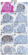
PC2 and 7B2 expression in rat pancreatic islet. (A) HE staining. (B) PC2 was expressed throughout the islets, with particularly robust expression in the periphery. (C) In ab22699 labeling, a similar pattern to that of PC2 was observed. (E) In #1–14 labeling, the immunoreaction was predominantly observed in the peripheral region. (G) In the instance of #77–90 labeling, the immunoreaction was observed to occur in a pervasive manner throughout the islet cells. (I) In #156–168 labeling, only a weak reaction was observed in the peripheral region. Negative controls were reacted with normal rabbit IgG (Rb-IgG) (D) or pre-absorbed antibodies with immunogen peptides, aa 1–14 (F), aa 77–90 (H), and aa 156–168 (J). Abbreviations: Is, islets; Ex, exocrine part. Scales, 50 µm.
Figure 5.
Figure 5.
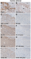
PNMT, PC2, and 7B2 expression in canine adrenal gland. (A) The presence of a positive PNMT immunoreaction in the medullary cells differentiates A cells (asterisk) from NA cells (double asterisks). (B) The PC2 immunoreaction exhibited variability among cells. For 7B2, a comparable immunoreactive pattern of was observed in the labeling of with ab22699 (C), #1–14 (E), #77–90 (G), and #156–168 (I). Negative controls were reacted with normal rabbit IgG (Rb-IgG) (D) or pre-absorbed antibodies with immunogen peptides, aa 1–14 (F), aa 77–90 (H), and aa 156–168 (J). Abbreviations: Co, adrenal cortex; Me, adrenal medulla. Scales, 100 µm.
Figure 6.
Figure 6.
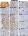
PNMT, PC2, and 7B2 expression in bovine adrenal gland. (A) Macroscopic transverse view of bovine adrenal gland. The outer and inner medullary zones are clearly distinguishable. (B) PNMT immunoreactivity was observed in the outer zone where A cells are concentrated. (C) PC2 expression was observed throughout the medulla and was particularly strong in some cells. (D and E) A positive immunoreaction of 7B2 labeled with ab22699 was observed with equal intensity in the outer and inner zones. (G and H) Immunoreaction labeled with #1–14 exhibited a stronger intensity in the outer zone compared with the inner zone. Furthermore, a subset of cells exhibited a discernible response, as indicated by the arrowheads. (J and K) A robust immunoreaction, identified through its labeling with #77–90, was observed in both the outer and inner zones, exhibiting equivalent intensity. (M and N) When labeled with #156–168, the overall reaction was weak, and some cells in the outer zone showed a moderate reaction (arrowheads). Negative controls were reacted with normal rabbit IgG (Rb-IgG) (F) or pre-absorbed antibodies with immunogen peptides, aa 1–14 (I), aa 77–90 (L), and aa 156–168 (O). Abbreviations: Co, adrenal cortex; Oz, outer zone of adrenal medulla; Iz, inner zone of adrenal medulla. Scales, 100 µm.
Figure 7.
Figure 7.
Reactivity of the 7B2 antibodies in porcine thyroid and chicken duodenum. HE staining of the porcine thyroid (A) and chicken duodenum (K) is shown. The presence of probable calcitonin-producing C cells in porcine thyroid and gastrointestinal endocrine cells in chicken duodenum is indicated by arrowheads. The expression of PC2 in probable C cells (B) and gastrointestinal endocrine cells (L) is also observed. The immunoreactive patterns of 7B2 labeled with ab22699 (C and M), #1–14 (E and O), #77–90 (G and Q), and #156–168 (I and S) were found to be similar. Negative controls included with normal rabbit IgG (Rb-IgG) (D and N) or pre-absorbed antibodies with immunogen peptides, aa 1–14 (F and P), aa 77–90 (H and R), and aa 156–168 (J and T). Abbreviations: Cr, crypt. Scales, 50 µm.
Figure 8.
Figure 8.
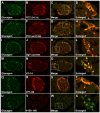
PC2 and 7B2 expression in α cells in rat islets. Double immunofluorescence labeling of glucagon (A, E, I, M, Q, and U), PC2 (D1E1S) (B), PC2 (ab191456) (F), 7B2 using ab22699 (J), #1–14 (N), #77–90 (R), and #156–168 (V) antibodies was performed. The immunoreactivity of PC2, which was labeled with D1E1S and ab191456, exhibited a comparable pattern, with pronounced expression in α cells situated within the islet periphery and minimal expression in the central region, where β cells are believed to be concentrated (C and G). (D and H) Enlarged panels of the rectangles in C and G, as well as putative β cells, are indicated by asterisks. A pattern of immunoreactivity of 7B2 in islets labeled with ab22699 (I–L) and #77–90 (Q–T) were found to be similar. However, immunoreactivity labeled with #1–14 exhibited variability in α cells and was not detected in the central β cell region (M–P). Furthermore, the immunoreactivity labeled with #156–168 in α cells was found to be minimal (U–X). Scales, 40 µm and 20 µm (enlarged images).
Figure 9.
Figure 9.
Coexpression of PC2 and 7B2 in rat and canine intermediate pituitary cells. Double immunofluorescence labeling technique was employed, utilizing an anti-PC2 (D1E1S) antibody in conjunction with anti-7B2 antibodies, #1–14, #77–90, and #156–168. This method was applied to the analysis of rat (A–I) and canine (J–R) pituitary glands. In the rat pituitary, all cells in the intermediate part exhibited PC2 expression (A, D, and G), and coexpression was pronounced in the labeling with #77–90 (D–F), in comparison to #1–14 (A–C) and #156–168 (G–I). A limited number of cells demonstrated immunoreactivity exclusively to 7B2 in the intermediate region (arrowheads). In the canine pituitary gland, a small number of PC2 and 7B2 coexpressed cells (thick arrows) were detected (J–L, M–O and P–R). The cells exhibiting only PC2 (thin arrows) or 7B2 (arrowheads) immunoreactivities were observed. In the #156-168 labeling, the signals were weak and punctate (Q). Abbreviations: Dis, distal part of the adenohypophysis; Int, intermediate part of the adenohypophysis; Neu, neurohypophysis. Scales, 40 µm.
Figure 10.
Figure 10.
Expression of PC2 and 7B2 in α-MSH-labeled canine pituitary melanotrophs. Double immunofluorescence labeling was utilized, employing anti-α-MSH antibody in conjunction with anti-PC2 (E-8), anti-7B2 (#1–14, #77–90, and #156–168) antibodies. The majority of cells in the intermediate part were positive for α-MSH expression (A, D, G, and J), and all PC2-positive cells coexpressed α-MSH (A–C) (thick yellow arrows). In addition, 7B2-positive cells demonstrated coexpression of α-MSH (D–L) (thick white arrows). Melanotrophs expressing only α-MSH were indicated by thin arrows, and a few cells in which 7B2 was detected with hardly detected α-MSH were indicated by arrowheads. The immunoreactivity of anti-PC2 antibodies, D1E1S and E-8, demonstrated consistent results (M–O). Scales, 40 µm.
Figure 11.
Figure 11.
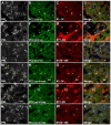
Coexpression of PC2 and 7B2 in rat pituitary mammotrophs and colocalization on granules. Triple immunofluorescence labeling utilized anti-PRL, anti-PC2 (D1E1S and ab191456), and anti-7B2 (#1–14, #77–90, and #156–168) antibodies. In mammotrophs, the immunoreactivity of PC2 labeled with D1E1S (B, F, and J) was weak compared with non-mammotrophs (B, F, and J). In contrast, the immunoreactivity labeled with ab191456 exhibited notable strength in mammotrophs (N, R, and V). The coexpression of PC2 and 7B2 in mammotrophs was prominent in combination with labeling of ab191456 and #1–14 (M–P) or ab191456 and #77–90 (Q–T) (thick yellow arrows). While a number of mammotrophs exhibited labeling with both D1E1S and #77–90 (E–H) (thick white arrows), a significant proportion of mammotrophs demonstrated minimal labeling with either D1E1S, #1–14 (A–D), or #77–90 (E–H) (white arrowheads). In non-mammotrophs, coexpression of PC2 and 7B2 labeled with D1E1S and #1–14 or with #77–90 was observed (asterisks); however, this was not observed with ab191456 (double asterisks). The presence of punctate signals designated as #156–168 was observed in mammotrophs (I–L and U–X) (thin white arrows), with no overlap observed between these signals and PC2. Scales, 10 µm.
Figure 12.
Figure 12.
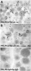
Triple immunogold labeling for PRL, PC2, and 7B2 in rat mammotrophs. Sections were subjected to incubation with primary antibody mixtures of anti-PRL, anti-PC2 (ab191456), and anti-7B2: (A) #1–14, or (B) #77–90. Colloidal gold-labeled secondary antibodies were utilized to detect mouse anti-PRL antibody (6 nm gold particles, small arrows), rabbit anti-PC2 antibody (18 nm gold particles, large arrows), and guinea pig anti-7B2 antibodies (12 nm gold particles, arrowheads). (C) In the negative control, normal rabbit IgG (Rb-IgG) and normal guinea pig IgG (Gp-IgG) were utilized in lieu of the anti-PC2 and anti-7B2 antibodies. Adjacent non-mammotrophic granules are indicated by asterisks. Scales, 100 nm.
Figure A1.
Figure A1.
Measurement of serum antibody titers. The antibody titers of antisera obtained by immunizing two guinea pigs (Gp1 and Gp2) with three types of 7B2 antigen peptides, A and A’ (aa1–14), B and B’ (aa77–90), and C and C’ (aa156–168), were measured using enzyme-linked immunosorbent assay (ELISA). The first group (Gp1) is displayed on the left side of the figure, while the second group (Gp2) is shown on the right. In comparison to the pre-immune serum, which is represented by a circle, high levels of absorption were observed at dilutions ranging from 8,000 to 16,000 times for both the sample collected for testing following three immunizations (illustrated as a diamond) and the sample obtained from the final whole blood collection (represented by a square).

References

    1. Hsi KL, Seidah NG, De Serres G, Chrétien M. Isolation and NH2-terminal sequence of a novel porcine anterior pituitary polypeptide. Homology to proinsulin, secretin and Rous sarcoma virus transforming protein TVFV60. FEBS Lett. 1982;147(2):261–6. - PubMed
    1. Seidah NG, Hsi KL, De Serres G, Rochemont J, Hamelin J, Antakly T, Cantin M, Chrétien M. Isolation and NH2-terminal sequence of a highly conserved human and porcine pituitary protein belonging to a new superfamily. Immunocytochemical localization in pars distalis and pars nervosa of the pituitary and in the supraoptic nucleus of the hypothalamus. Arch Biochem Biophys. 1983;225(2):525–34. - PubMed
    1. Iguchi H, Chan JS, Seidah NG, Chrétien M. Tissue distribution and molecular forms of a novel pituitary protein in the rat. Neuroendocrinology. 1984;39(5):453–8. - PubMed
    1. Taupenot L, Harper KL, O’Connor DT. The chromogranin-secretogranin family. N Engl J Med. 2003;348(12):1134–49. - PubMed
    1. Bartolomucci A, Possenti R, Mahata SK, Fischer-Colbrie R, Loh YP, Salton SR. The extended granin family: structure, function, and biomedical implications. Endocr Rev. 2011;32(6):755–97. - PMC - PubMed

LinkOut - more resources