

Current Insights and Management Strategies for Lower Cervical Arteriovenous Fistulas: A Comprehensive Review
- PMID: 40852074
- PMCID: PMC12370346
- DOI: 10.1055/s-0045-1809046
Current Insights and Management Strategies for Lower Cervical Arteriovenous Fistulas: A Comprehensive Review
Abstract
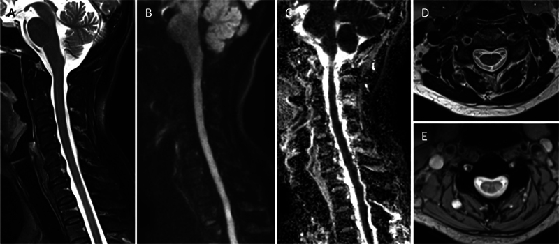
Lower cervical arteriovenous fistulas (AVFs) are rare and complex vascular malformations that pose significant clinical challenges due to their location and variable presentation. While upper cervical AVFs have been extensively studied, lower cervical AVFs remain underresearched. This study aims to review the clinical presentations, management strategies, and outcomes of patients with lower cervical AVFs to enhance understanding and improve treatment approaches. We conducted a retrospective analysis of patients with spinal vascular malformations treated at our institute between June 2006 and December 2023, identifying two cases of lower cervical AVFs. Additionally, a systematic literature review was performed following the Preferred Reporting Items for Systematic Reviews and Meta-Analyses guidelines, including 44 patients with lower cervical AVFs, using databases such as Ovid MEDLINE, PubMed, and Cochrane. Data collected included patient demographics, clinical presentation, fistula type, arterial and venous involvement, treatment modality, and neurological outcomes. Among the 44 patients with lower cervical AVFs, including our two cases, 50% were female, and the mean age was 48.68 years (range: 4-76 years). Clinical presentations varied, with 27.3% experiencing hemorrhage, 18.2% presenting with myelopathy, and 18.2% remaining asymptomatic. Venous drainage patterns played a significant role in symptom severity, with complex perimedullary and retrograde venous drainage contributing to worse outcomes. Treatment included endovascular embolization (40.9%), surgical resection (25%), and combined approaches (18.2%), with good recovery achieved in 54.5% of cases. Lower cervical AVFs present diverse clinical challenges due to their variable presentations and complex vascular anatomy. Early diagnosis and tailored management, including endovascular embolization and surgical resection, are essential for optimizing patient outcomes. Further research is needed to better understand the natural history of asymptomatic AVFs and improve treatment protocols.
Keywords: epidural arteriovenous fistulas; lower cervical arteriovenous fistulas; perimedullary arteriovenous fistulas; radicular arteriovenous fistulas; spinal cord venous infarction; spinal dural arteriovenous fistula.
Asian Congress of Neurological Surgeons. This is an open access article published by Thieme under the terms of the Creative Commons Attribution-NonDerivative-NonCommercial License, permitting copying and reproduction so long as the original work is given appropriate credit. Contents may not be used for commercial purposes, or adapted, remixed, transformed or built upon. ( https://creativecommons.org/licenses/by-nc-nd/4.0/ ).
Conflict of interest statement
Conflict of Interest None declared.
Figures


Similar articles
-
Comparative analysis of spinal extradural arteriovenous fistulas with or without intradural venous drainage: a systematic literature review.Neurosurg Focus. 2012 May;32(5):E8. doi: 10.3171/2012.2.FOCUS1216. Neurosurg Focus. 2012. PMID: 22537134
-
Angioarchitecture Classification and Treatment Modalities of Craniocervical Junction Arteriovenous Fistulas: A Cohort Study of 155 Patients.Neurosurgery. 2024 Sep 1;95(3):692-701. doi: 10.1227/neu.0000000000002939. Epub 2024 Apr 15. Neurosurgery. 2024. PMID: 39145652
-
Spinal dural arteriovenous fistulas presenting as intracranial subarachnoid hemorrhage: A systematic review.Interv Neuroradiol. 2025 Jul 15:15910199251328721. doi: 10.1177/15910199251328721. Online ahead of print. Interv Neuroradiol. 2025. PMID: 40660916 Free PMC article. Review.
-
Dural arteriovenous fistulas of the hypoglossal canal: systematic review on imaging anatomy, clinical findings, and endovascular management.J Neurosurg. 2015 Apr;122(4):883-903. doi: 10.3171/2014.10.JNS14377. Epub 2014 Nov 21. J Neurosurg. 2015. PMID: 25415064
-
Endovascular Treatment for Tentorial Dural Arteriovenous Fistulas: A Retrospective Single-Center Study.AJNR Am J Neuroradiol. 2025 Aug 1;46(8):1617-1624. doi: 10.3174/ajnr.A8676. AJNR Am J Neuroradiol. 2025. PMID: 39880688
References
-
- Iampreechakul P, Wangtanaphat K, Wattanasen Y, Hangsapruek S, Lertbutsayanukul P, Siriwimonmas S. Dural arteriovenous fistula of the craniocervical junction along the first cervical nerve: a single-center experience and review of the literature. Clin Neurol Neurosurg. 2023;224:107548. - PubMed
-
- Li J, Lin F, Zhu J et al. Enhanced treatment options for dural arteriovenous fistulas at the craniocervical junction: endovascular embolization versus microsurgery? A single-center 23-year experience. World Neurosurg. 2024;182:e414–e430. - PubMed
-
- Willinsky R, TerBrugge K, Lasjaunias P, Montanera W. The variable presentations of craniocervical and cervical dural arteriovenous malformations. Surg Neurol. 1990;34(02):118–123. - PubMed
Publication types
LinkOut - more resources
Full Text Sources