. 2025 Aug;7(8):2400867.
doi: 10.1002/aisy.202400867. Epub 2025 Apr 15.

High-Throughput Nanorheology of Living Cells Powered by Supervised Machine Learning

Affiliations

High-Throughput Nanorheology of Living Cells Powered by Supervised Machine Learning

Jaime R Tejedor et al. Adv Intell Syst. 2025 Aug.

Abstract

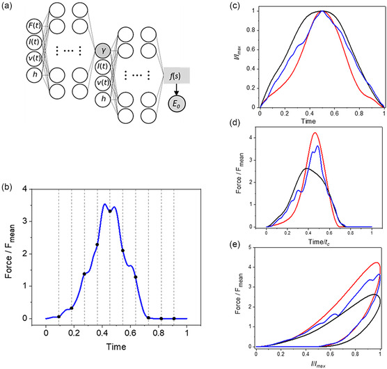
Atomic force microscopy (AFM) is extensively applied to measure the nanomechanical properties of living cells. Despite its popularity, some applications on mechanobiology are limited by the low throughput of the technique. Currently, the analysis of AFM-nanoindentation data is performed by model fitting. Model fitting is slow, data intensive, and prone to error. Herein, a supervised machine-learning regressor is developed for transforming AFM force-distance curves into nanorheological behavior. The method reduces the computational time required to process a force volume of a cell made of 2.62 × 10⁵ curves from several hours to minutes. In fact, the regressor increases the throughput by 50-fold. The training and the validation of the regressor are performed by using theoretical curves derived from a contact mechanics model that combined power-law rheology with bottom effect corrections and functional data analysis. The regressor predicts the modulus and the fluidity coefficient of mammalian cells with a relative error below 4%.

Keywords: atomic force microscopies; mammalian cells; mechanobiologies; nanoindentations; nanorheologies.


Conflict of interest statement

The authors declare no conflict of interest.

Figures

Figure 1
Figure 1
a) Scheme of an AFM‐nanoindentation experiment on a cell. b) Examples of some common indentation profiles: triangular and capped sinusoidal indentation profiles (v = 10 μm s−1). c) Synthetic (theoretical) force versus time curves for the indentations shown in (b). d) FDCs obtained by combining (b) and (c). The synthetic FDCs were calculated by using a cell with E 0 = 5 kPa, γ = 0.3, and h = 7 μm. e,f) Experimental FDCs (black) and single power‐law rheology fittings (red). The FDCs were obtained at (e) 10 μm s−1 and (f) 300 μm s−1 on an HeLa cell (over the nuclear region). The modulus and fluidity parameters are included in the panels. Experimental FDCs by Gisbert et al.[ 14 ]
Figure 2
Figure 2
a) Scheme of the SML regressor. The first neural network predicts the fluidity coefficient and the second neural network predicts the mean value of the dimensionless force, f(s). The modulus is obtained from that value (Equation 15). b) The force is uniformly sampled in the time domain to provide a discretized set of input values. The regressor is trained by sampling 100 time points and a base of splines with 40 knots and degree 3. c) Some examples of the indentation profiles used in training. d) Force–time curves for the indentation profiles shown in (c). e) FDCs for the profiles shown in (c). Parameters: γ = 0.3, I max = 0.5 h, and p = 0.5.
Figure 3
Figure 3
Validation test. Comparison between predicted (regressor) and true (synthetic data). The regressor predicts the modulus and the fluidity coefficient, respectively, with a relative error below 2% and 0.5%. Notice that the same regressor predicts the properties of very stiff (100 kPa) and very soft cells (0.1 kPa). Those values cover the range of values measured on living mammalian cells. The range fluidity coefficient values applied to train the regressor covered all possible values for a material, from an elastic solid (γ = 0) to a Newtonian liquid (γ = 1).
Figure 4
Figure 4
a) Predicted nanorheology of HeLa cells by the SML regressor. b) Predicted nanorheology of an NIH 3T3 fibroblast cell. c) Relation fluidity coefficient and modulus (HeLa). d) Relation fluidity coefficient and modulus (NIH 3T3 fibroblast). The experimental FDCs were obtained by probing the cells at different velocities, for this reason, a color‐code scale bar was included. Experimental FDCs were obtained from an article.[ 14 ]
Figure 5
Figure 5
a) High‐spatial resolution compositional map of an HeLa cell powered by machine learning. The map was obtained by applying the regressor to AFM experimental data. The map combined b) topography and c) modulus data. The compositional map shows the fine structure of the actin filament network, the local variation of the modulus, and the size and shape of the nucleus. (b) AFM topography. (c) Scaling modulus map. The final spatial resolution of the regressor map was limited by the number of data points (512 × 512). The experimental FDCs were obtained with a JPK NanoWizard 3.[ 14 ]

Similar articles

Cited by

References

    1. Efremov Y. M., Okajima T., Raman A., Soft Matter 2020, 16, 64. - PubMed
    1. Garcia R., Chem. Soc. Rev. 2020, 49, 5850.
    1. Flormann D. A. D., Anton C., Pohland M. O., Bautz Y., Kaub K., Terriac E., Schäffer T. E., Rheinlaender J., Janshoff A., Ott A., Lautenschläger F., Front. Phys. 2021, 9, 711860.
    1. Solares S. D., Cartagena‐Rivera A. X., Beilstein J. Nanotechnol. 2022, 13, 1483. - PMC - PubMed
    1. Li M., Liu L., Xiao X., Xi N., Wang Y., IEEE Trans. Nanobiosci. 2016, 15, 398.

LinkOut - more resources