. 2025 May 15;4(2):22.
doi: 10.20517/mrr.2025.10. eCollection 2025.

Understanding the interaction between melatonin and bifidobacteria

Affiliations

Understanding the interaction between melatonin and bifidobacteria

Sonia Mirjam Rizzo et al. Microbiome Res Rep. .

Abstract

Aim: The human gastrointestinal tract is home to a complex and dynamic microbial community, known as the gut microbiota, which begins to form at birth and evolves throughout life. Among the factors influencing its initial development, breastfeeding is one of the most important. Human milk is a chemically complex body fluid, including hormones, like melatonin, which is involved in regulating the sleep-wake cycle, helping to establish the newborn's circadian rhythm. In the current study, the molecular interactions between human melatonin and a bifidobacteria-rich infant gut microbiota were explored. Methods: Possible molecular communication was assessed using in vitro assays and functional genomic approaches. Results: Our results highlight that melatonin elicits different functional microbial impacts, both at transcriptional and phenotypic levels (i.e., adhesion to intestinal cells), that are dependent on the bifidobacterial species analyzed. Conclusion: Among the bifidobacterial taxa assayed, Bifidobacterium bifidum demonstrated the highest level of molecular interaction with melatonin, highlighting its significant role in this process.

Keywords: CST; Melatonin; bifidobacteria; gut microbes; hormones.


Conflict of interest statement

Longhi G serves as Junior Editorial Board member, van Sinderen D as Co-Editor-in-Chief, Ventura M as Editor-in-Chief, and Turroni F as Executive Editor of Microbiome Research Reports. None of these individuals was involved in any aspect of the editorial process for this manuscript, including reviewer selection, manuscript handling, or decision making. The other authors declared that there are no conflicts of interest.

Figures

Figure 1
Figure 1
Effects of melatonin on B. bifidum PRL2010 transcriptomes. (A) Displays a Venn diagram illustrating the number of shared and unique B. bifidum PRL2010 upregulated genes, when exposed alone (on the left) or with other microorganisms representing the ICST-BI/EN (on the right), in the presence of melatonin hormone vs. control; (B) Depicts the statistically significantly expressed B. bifidum PRL2010 genes (with logFC in transcription ≥ 1 in combination with a P-value ≤ 0.05) encoding extracellular structures and transporters possibly related to the interaction with the host. logFC: log2 fold change.
Figure 2
Figure 2
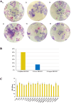
Adherence of B. bifidum PRL2010, B. longum PRL2022, and B. breve PRL2012 to HT29-MTX cells monolayers and growth assay of B. bifidum PRL2010 in the presence of different concentrations of melatonin. (A) Displays the light microscopic images of HT29-MTX monolayers as observed with Giemsa staining of bifidobacterial species grown in the absence (1, 3, 5) or in the presence of melatonin (2, 4, 6), respectively. The bifidobacterial species shown in each image are: (1, 2) B. bifidum PRL2010, B. breve PRL2022 (3, 4), and B. longum PRL2012 (5, 6); (B) Depicts the percentage increase in adhesion capacity of B. bifidum PRL2010, B. longum PRL2022, and B. breve PRL2022 when in the presence of melatonin, compared to their adhesion in the absence of the hormone; (C) Represents the growth assay of B. bifidum PRL2010 in the presence of different amounts of melatonin. Each pillar shows the bacterial growth at the 19 selected melatonin concentrations from 8 µM to 58.59 pM or under control conditions (CTRL, growth of the bifidobacteria without the hormone). The OD600nm values were expressed as the average of three independent replicates. No statistical significances were exhibited between the conditions, P-value > 0.

References

    1. Jandhyala SM, Talukdar R, Subramanyam C, Vuyyuru H, Sasikala M, Nageshwar Reddy D. Role of the normal gut microbiota. World J Gastroenterol. 2015;21:8787–803. doi: 10.3748/wjg.v21.i29.8787. - DOI - PMC - PubMed
    1. Hill CJ, Lynch DB, Murphy K, et al. Evolution of gut microbiota composition from birth to 24 weeks in the INFANTMET Cohort. Microbiome. 2017;5:4. doi: 10.1186/s40168-016-0213-y. - DOI - PMC - PubMed
    1. Vandenplas Y, Carnielli VP, Ksiazyk J, et al. Factors affecting early-life intestinal microbiota development. Nutrition. 2020;78:110812. doi: 10.1016/j.nut.2020.110812. - DOI - PubMed
    1. Rizzo SM, Alessandri G, Lugli GA, et al. Exploring molecular interactions between human milk hormone insulin and bifidobacteria. Microbiol Spectr. 2023;11:e0066523. doi: 10.1128/spectrum.00665-23. - DOI - PMC - PubMed
    1. Korpela K, Blakstad EW, Moltu SJ, et al. Intestinal microbiota development and gestational age in preterm neonates. Sci Rep. 2018;8:2453. doi: 10.1038/s41598-018-20827-x. - DOI - PMC - PubMed

LinkOut - more resources