. 2025 Aug 11;10(32):36178-36202.
doi: 10.1021/acsomega.5c03992. eCollection 2025 Aug 19.

Hepatoprotective Potential of Green Synthesized Nanoparticles from Peel Extract against HFS Diet-Induced NASH in Mice, Integrated with Chemical Profiling and Molecular Modeling

Affiliations

Hepatoprotective Potential of Green Synthesized Nanoparticles from Peel Extract against HFS Diet-Induced NASH in Mice, Integrated with Chemical Profiling and Molecular Modeling

Sameh S Elhady et al. ACS Omega. .

Abstract

is known for its wide variety of secondary metabolites, including essential oil, alkaloids, flavonoids, and phenolic acids, which are considered the reason for its diverse potential medical uses. Herein, extract was prepared using different formulas of metal oxide nanoparticles as ZnO NPs and MgO NPs with 22.5 ± 1.3 and 18.3 ± 1.5 nm average particle sizes, respectively. The protective effects of extract and its two nano formulas against NASH induced by a high-fat, high-sucrose (HFS) diet in mice were evaluated. The NASH mice showed hepatic steatosis, inflammation, reduced liver function, increased body, liver, and fat weights, elevated hepatic index, and disrupted serum lipid profiles. Additionally, the mice displayed heightened hepatic oxidative stress and increased expression of inflammatory and profibrotic markers, along with altered lipid metabolism, indicated by elevated levels of SIRT-1, FGF-21, and SREBP-1c. Notably, treatment with extract and its metallic nanoparticle formulations alleviated these abnormalities, with the most significant improvement observed in the MgO NP formulation. Finally, extract was subjected to a deep phytochemical screening through Liquid chromatography combined with mass spectrometry (LC-MS/MS) analysis, revealing the presence of a high diversity of polyphenolics that may contribute to the suggested therapeutic effects of the crude extract. A molecular docking study highlighted the binding affinities of all identified flavonoid compounds, especially Quercetin, toward IL-1β, TNF-α, and TGF-β target proteins. This shows good translation for the cumulative anti-inflammatory effect depicted by the crude extract. Thermodynamic stability of Quercetin toward each bound proinflammatory protein was confirmed through 150 ns all-atom molecular dynamics simulations. Overall, current findings suggest that exerts a protective effect against NASH, which could be enhanced by nano formulas.


Figures

1
1
Chemical structure of identified compounds in extract through LC–MS/MS.
1
1
Chemical structure of identified compounds in extract through LC–MS/MS.
2
2
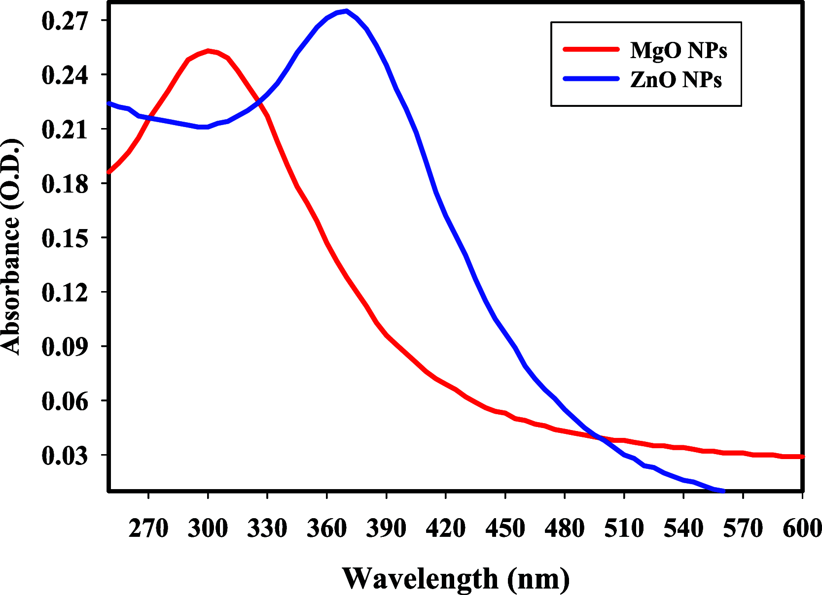
UV–vis spectra of the synthesized MgO, and ZnO NPs (diluted 10 times).
3
3
Size distribution of the synthesized ZnO (a), and MgO (b) nanoconjugates by DLS analysis.
4
4
XRD spectra of the synthesized MgO, and ZnO nanoconjugates.
5
5
Effect of extract and its metallic nanoparticles formulas on the hepatic histopathological changes and NASH grading. NASH, nonalcoholic steatohepatitis. The normal group showed uniform liver tissue, showing regular cords of hepatocytes (Black arrows). In NASH group, there is an obvious steatosis and hydropic degeneration in several zone 3 hepatocytes (Black arrows). There are foci of intralobular inflammation by chronic inflammatory cells (Black arrowheads). No portal tracts inflammation is seen. (150 mg/kg) group also showed obvious steatosis and hydropic degeneration in several zone 3 hepatocytes (Black arrows). There are no detected foci of intralobular inflammation. No portal tracts inflammation is seen. MgO and ZnO NPs- groups showed significant improvement in hepatocytes which show mild hydropic degeneration and steatosis (Black arrows). There is no detected intralobular or portal tracts inflammation (Black arrowhead) (H&E, 20x). Data of the NASH grading are presented as % of affected animals (n = 8).
6
6
Effect of extract and its metallic nanoparticles formulas on the serum activities of (A) ALT and (B) AST. NASH, nonalcoholic steatohepatitis; ALT, alanine aminotransferase; AST, aspartate aminotransferase. Data are displayed as mean ± SD and analyzed using one-way ANOVA followed by Tukey’s post hoc test (n = 8). (a) Indicates significant difference from the normal control group; (b) indicates a significant difference from the NASH group; (c) indicates a significant difference from the (150 mg/kg) group; (d) indicates a significant difference from the ZnO NPs- (40 mg/kg) group. p < 0.05 was indicative of a significant difference.
7
7
Effect of extract and its metallic nanoparticles formulas on (A) final body weight, (B) body weight over the 12 week experimental period, (C) liver weight, (D) liver index, and (E) fat weight around epididymis. NASH, nonalcoholic steatohepatitis. Data are displayed as mean ± SD and analyzed using one-way ANOVA followed by Tukey’s post hoc test (n = 8). (a) Indicates a significant difference from the normal control group; (b) indicates a significant difference from the NASH group; (c) indicates a significant difference from the (150 mg/kg) group; (d) indicates a significant difference from the ZnO NPs- (40 mg/kg) group. p < 0.05 was indicative of a significant difference.
8
8
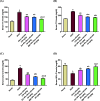
Effect of extract and its metallic nanoparticle formulas on the serum levels of (A) TC, (B) TG, (C) LDL-C, and (D) HDL-C. NASH, nonalcoholic steatohepatitis; TC, total cholesterol; TG, triglyceride; LDL-C, low-density lipoprotein cholesterol; HDL-C, high-density lipoprotein cholesterol. Data are displayed as mean ± SD and analyzed using one-way ANOVA followed by Tukey’s post hoc test (n = 8). (a) Indicates a significant difference from the normal control group; (b) indicates a significant difference from the NASH group; (c) indicates a significant difference from the (150 mg/kg) group; (d) indicates a significant difference from the ZnO NPs- (40 mg/kg) group. p < 0.05 was indicative of a significant difference.
9
9
Effect of extract and its metallic nanoparticle formulas on the hepatic tissue levels of (A) MDA and (B) TAC. NASH, nonalcoholic steatohepatitis; MDA, malondialdehyde; TAC, total antioxidant capacity. Data are displayed as mean ± SD and analyzed using one-way ANOVA followed by Tukey’s post hoc test (n = 8). (a) Indicates a significant difference from the normal control group; (b) indicates a significant difference from the NASH group; (c) indicates a significant difference from the (150 mg/kg) group; (d) indicates a significant difference from the ZnO NPs- (40 mg/kg) group. p < 0.01 was indicative of a significant difference.
10
10
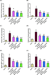
Effect of extract and its metallic nanoparticle formulas on the hepatic tissue gene expression of (A) TLR-4, (B) NF-κB, (C) TNF-α (D) IL-1β, (E) MCP-1, and (F) TGF-β. NASH, nonalcoholic steatohepatitis; TLR-4, toll-like receptor-4; NF-κB, nuclear factor kappa B; TNF-α, tumor necrosis factor-alpha; IL-1β, interleukin-1 beta; MCP-1, monocyte chemoattractant protein-1; TGF-β, transforming growth factor-beta. Data are displayed as mean ± SD and analyzed using one-way ANOVA followed by Tukey’s post hoc test (n = 8). (a) Indicates a significant difference from the normal control group; (b) indicates a significant difference from the NASH group; (c) indicates a significant difference from the (150 mg/kg) group; (d) indicates a significant difference from the ZnO NPs- (40 mg/kg) group. p < 0.05 was indicative of a significant difference.
11
11
Effect of extract and its metallic nanoparticle formulas on the hepatic tissue levels of (A) SIRT-1, (B) FGF-21, and (C) SREBP-1c. NASH, nonalcoholic steatohepatitis; SIRT-1, sirtuin 1; FGF-21, fibroblast growth factor-21; SREBP-1c, sterol regulatory element binding protein-1c. Data are displayed as mean ± SD and analyzed using one-way ANOVA followed by Tukey’s post hoc test (n = 8). (a) Indicates a significant difference from the normal control group; (b) indicates a significant difference from the NASH group; (c) indicates a significant difference from the (150 mg/kg) group; (d) indicates a significant difference from the ZnO NPs- (40 mg/kg) group. p < 0.05 was indicative of a significant difference.
12
12
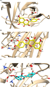
Binding disposition of Quercetin toward the tested proteins of IL-1β (A), TNF-α (B), and TGF-β (C) highlights the binding interactions with the key amino acids inside protein active sites. The 3-D images were generated using Chimera-UCSF. A full report of binding interactions of all flavenoid structures is summarized in the supplementary file (Table S1).
13
13
Explicit molecular dynamics simulated Quercetin at the three proinflammatory targets. (A) C-alpha RMSDs of proinflammatory proteins and RMSDs of individual Quercetin; (B) Overlaid extracted frames at beginning (0 ns), midway (75 ns), and end (150 ns) of the simulation runs. Proteins as cartoons and ligand as sticks are colored in respect to extracted frames; 0, 75, and 150 ns → green, yellow, and red; (C) total free binding energies and respective contributing energy terms; (D) Residue-wise energy contributions of the proteins target canonical pocket.
13
13
Explicit molecular dynamics simulated Quercetin at the three proinflammatory targets. (A) C-alpha RMSDs of proinflammatory proteins and RMSDs of individual Quercetin; (B) Overlaid extracted frames at beginning (0 ns), midway (75 ns), and end (150 ns) of the simulation runs. Proteins as cartoons and ligand as sticks are colored in respect to extracted frames; 0, 75, and 150 ns → green, yellow, and red; (C) total free binding energies and respective contributing energy terms; (D) Residue-wise energy contributions of the proteins target canonical pocket.

Similar articles

References

    1. Prakash S., Rai U., Kosuru R., Tiwari V., Singh S.. Amelioration of diet-induced metabolic syndrome and fatty liver with sitagliptin via regulation of adipose tissue inflammation and hepatic Adiponectin/AMPK levels in mice. Biochimie. 2020;168:198–209. doi: 10.1016/j.biochi.2019.11.005. - DOI - PubMed
    1. Teng W., Zhao L., Yang S., Zhang C., Liu M., Luo J., Jin J., Zhang M., Bao C., Li D.. et al. The hepatic-targeted, resveratrol loaded nanoparticles for relief of high fat diet-induced nonalcoholic fatty liver disease. J. Controlled Release. 2019;307:139–149. doi: 10.1016/j.jconrel.2019.06.023. - DOI - PubMed
    1. Zhang R., Chu K., Zhao N., Wu J., Ma L., Zhu C., Chen X., Wei G., Liao M.. Corilagin Alleviates Nonalcoholic Fatty Liver Disease in High-Fat Diet-Induced C57BL/6 Mice by Ameliorating Oxidative Stress and Restoring Autophagic Flux. Front. Pharmacol. 2020;10:1693. doi: 10.3389/fphar.2019.01693. - DOI - PMC - PubMed
    1. Ore A., Ugbaja R. N., Adeogun A. I., Akinloye O. A.. An albino mouse model of nonalcoholic fatty liver disease induced using high-fat liquid “Lieber-DeCarli” diet: a preliminary investigation. Porto Biomed. J. 2020;5(4):e071. doi: 10.1097/j.pbj.0000000000000071. - DOI - PMC - PubMed
    1. Tarantino G., Citro V., Balsano C.. Liver-spleen axis in nonalcoholic fatty liver disease. Expert Rev. Gastroenterol. Hepatol. 2021;15(7):759–769. doi: 10.1080/17474124.2021.1914587. - DOI - PubMed

LinkOut - more resources