. 2025 Dec;16(1):2546068.
doi: 10.1080/21505594.2025.2546068. Epub 2025 Aug 25.

MoMkt1, a member of XPG/RAD2 nuclease family, regulates development and pathogenicity in Magnaporthe oryzae

Affiliations

MoMkt1, a member of XPG/RAD2 nuclease family, regulates development and pathogenicity in Magnaporthe oryzae

Na Li et al. Virulence. 2025 Dec.

Abstract

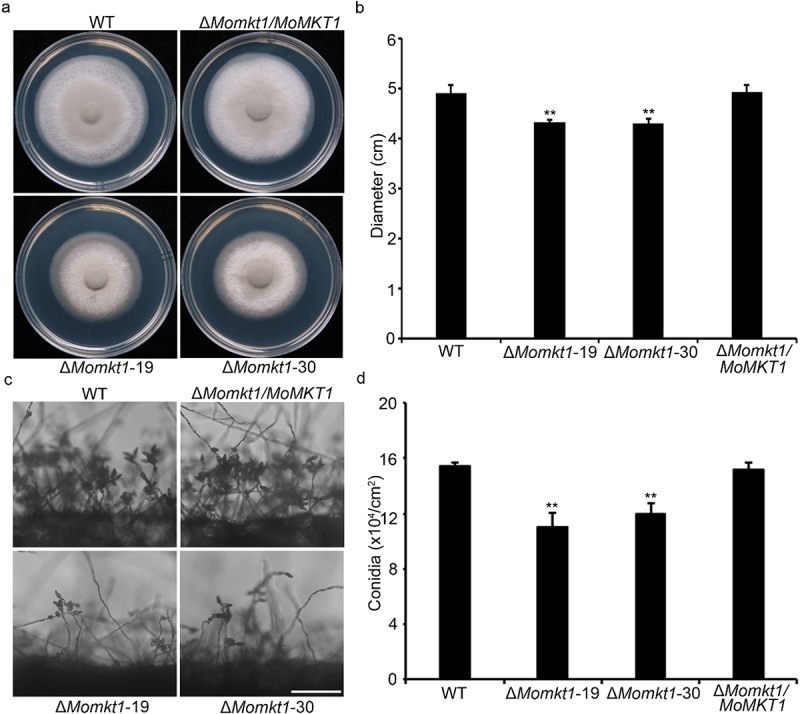
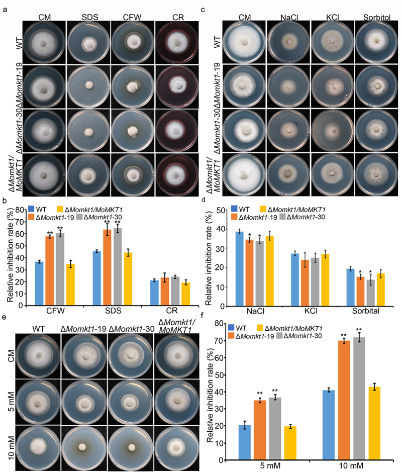
XPG/RAD2 nuclease family plays a crucial role in DNA damage repair to maintain genomic integrity. However, the biological function of Mkt1, a member of the XPG/RAD2 nuclease family, remains unclear in Magnaporthe oryzae. In this study, we identified and characterized the biological functions of MoMkt1. Our results demonstrated that MoMkt1 is involved in hyphal growth, conidiation, normal appressorium development, degradation of glycogen and lipid droplets, cell wall stress and oxidative stress responses, and pathogenicity in M. oryzae. Additionally, MoMkt1 is required for scavenging host-derived reactive oxygen species (ROS) and responding to DNA replicative stress. Further investigation revealed that MoMkt1 interacts with the transcription factor IIH (TFIIH) core subunit MoTfb2. Overexpression (OE) of MoTFB2 slightly affected the virulence of M. oryzae. Transcriptome data revealed that MoMKT1 regulates metabolic pathways, cell wall, oxidation-reduction processes, and DNA repair. Our study provides insights into MoMkt1-mediated development and pathogenicity in M. oryzae.

Keywords: DNA repair; Magnaporthe oryzae; MoMkt1; XPG/RAD2 nuclease family; pathogenicity.


Conflict of interest statement

No potential conflict of interest was reported by the author(s).

Figures

Figure 1.
Figure 1.
Characterization and subcellular localization of MoMkt1 protein in M. oryzae. (a) Phylogenetic analysis of MoMkt1 and its homologs from different organisms using the neighbor-joining method by MEGA software. GenBank accession numbers and the corresponding species names are as follows: XP_003715774.1 (Magnaporthe oryzae); XP_009223415.1 (Gaeumannomyces tritici); XP_960742.1 (Neurospora crassa); KAI6766631 (Fusarium graminearum); XP_024550529.1 (Botrytis cinerea); XP_050469344.1 (Aspergillus nidulans); XP_567528.1 (Cryptococcus neoformans); NP_174256.2 (Arabidopsis thaliana); KAI4085534.1 (Homo sapiens); NP_036142.2 (Mus musculus); NP_014314.3 (Saccharomyces cerevisiae). (b) Protein domain analysis of Mkt1 was performed using pfam website (https://www.ebi.ac.uk/interpro/). The amino acid (aa) residue number at the start and end of the protein, as well as the predicted domains, were labeled at the top or bottom of the protein. (c) Subcellular localization distribution of MoMkt1 at different developmental stages. Transformant expressing MoMkt1-GFP and H1-RFP plasmids in the WT strain Guy11 was observed using a laser scanning confocal microscope (Nikon, Japan). HY, hypha; CO, Conidium; AP, appressorium at 6 hpi; IH, invasive hyphae at 36 hpi. Bar, 20 μm.
Figure 2.
Figure 2.
MoMKT1 is involved in the vegetative growth and conidiation of M. oryzae. (a) Colonies of the WT strain Guy11, ΔMomkt1 mutants, and the complemented strain ΔMomkt1/MoMKT1 on CM plates were observed after 7 days at 28°C. (b) Statistical analysis of the colony diameter of the indicated strains. (c) Conidiophores of the indicated strains. Bar, 100 µm. (d) Statistical analysis of conidiation of the indicated strains. Error bars represent SD and asterisks indicate significant differences between the WT strain Guy11 and ΔMomkt1 mutants, estimated using the Student’s t-test (**, p < 0.01).
Figure 3.
Figure 3.
MoMKT1 is important for full virulence of M. oryzae. (a) Pathogenicity was tested on unwounded (U) and wounded (W) barley leaves. The mycelial agar plugs of the indicated strains were inoculated on 7-day-old barley leaves and photographed at 5 dpi. Bar, 10 mm. (b) Statistical analysis of the lesion area of the indicated strains on barley leaves using the ImageJ software. Error bars represent SD and asterisks indicate significant differences (**, p < 0.01). (c) 14-day-old rice seedlings were inoculated by spraying with 10 mL conidial suspensions (5 × 104 conidia/mL in a 0.2% (w/v) gelatin solution) from the indicated strains, and photographed at 5 dpi. Bar, 10 mm. (d) Statistical analysis of the number of lesions in the indicated strains within a 4 cm long leaf, and at least three leaves were counted for each strain. Three experiments were conducted. Error bars represent SD and asterisks indicate significant differences (**, p < 0.01).
Figure 4.
Figure 4.
MoMKT1 is important for appressorium formation, invasive hyphal expansion, and scavenging host ROS. (a) Appressorium formation in the WT strain Guy11, ΔMomkt1 mutants, and the complemented strain ΔMomkt1/MoMKT1 was induced on an artificial hydrophobic surface, and observed at 4, 6, 8, 12, and 24 hpi. Bar, 50 µm. (b) Statistical analysis of the appressorium formation rates of the tested strains. A total of 100 conidia were counted for each strain and three experiments were performed. (c) Four types of IH of M. oryzae in barley epidermal cells. Type 1 (T1), no penetration; type 2 (T2), single infectious hypha; type 3 (T3), more than two IH limited to one cell; type 4 (T4). IH extended to the adjacent cells. Bar, 50 μm. (d) Statistical analysis of each type IH of the indicated strains. A total of 100 infectious penetration sites were counted for each strain, and the experiment was repeated thrice. (e) DAB staining was performed on the infected barley leaves of the indicated strains at 30 hpi. Bar, 50 μm. (f) Statistical analysis of DAB-stained infected barley cells. For each strain, at least 100 infected barley cells were observed and three experiments were performed. (g) Barley leaves were inoculated with conidial suspensions of all tested strains treated with DPI, and invasive hyphae were observed at 30 hpi. The dimethyl sulfoxide (DMSO) was used as a control to dissolve the DPI. Bar, 50 μm. (h) Transcription level of ROS-detoxification‐related genes in the indicated strains. The β‐tubulin gene (MGG_00604) was used as the reference gene. Three independent biological experiments were conducted. Error bars represent SD and asterisks indicate significant differences (**, p < 0.01; *, p < 0.05). “NS” represents non-significant differences (NS, p > 0.05).
Figure 5.
Figure 5.
Disruption of MoMkt1 delays the mobilization and degradation of glycogen and lipid droplets. (a) Distribution of lipid droplets in conidia and appressorium of all tested strains stained with Nile red solution was observed under a light microscope at 0, 2, 8, 16 and 24 hours, respectively. Bar, 20 µm. (b) Statistical analysis of appressorium-containing lipid droplets in all tested strains. In total, 100 appressoria were counted. (c) Statistical analysis of conidia containing lipid droplets in all tested strains. A total of 100 conidia were counted in each experiment. (d) Distribution of glycogen in the conidia and appressorium of all tested strains stained with KI/I2 solution was observed under a light microscope at 0, 2, 8, 16, and 24 hours, respectively. Bar, 50 µm. (e) Statistical analysis of appressorium-containing glycogen in all the tested strains. In total, 100 appressoria were counted. Three independent biological experiments were conducted. (f) Statistical analysis of glycogen-containing conidia in all the tested strains. A total of 100 conidia were counted for each experiment. Error bars represent SD and asterisks indicate significant differences (**, p < 0.01; *, p < 0.05).
Figure 6.
Figure 6.
MoMkt1 is required for the cell wall stress response. (a) Colony morphology of the WT strain Guy11, ΔMomkt1 mutants, and the complemented strain ∆Momkt1/MoMKT1 were cultured on CM containing 200 µg/mL CFW, 0.01% SDS, and 600 µg/mL CR. The colonies were measured and photographed under dark conditions at 28°C for 7 days. (b) Statistical analysis of the relative inhibition rates (%) of the indicated strains. (c) Colony morphology of the tested strains on CM containing 0.7 M NaCl, 0.6 M KCl, and 1 M sorbitol in the dark at 28°C for 7 days. (d) Statistical analysis of the relative inhibition rates (%) of the indicated strains. (e) Colony morphology of the tested strains on CM containing 5 mM H2O2 and 10 mM H2O2 in the dark at 28°C for 7 days. (f) Statistical analysis of the relative inhibition rates (%) of the indicated strains. For each strain, three independent biological experiments were performed, with three replicates each time. Error bars represent SD and asterisks indicate significant differences (**, p < 0.01; * p < 0.05). “NS” represents non-significant differences (NS, p > 0.05).
Figure 7.
Figure 7.
MoMkt1 is involved in DNA replicative stress response. (a) Colony morphology of the tested strains on CM containing 10 mM HU in the dark at 28°C for 7 days. (b) Statistical analysis of the relative inhibition rates (%) of the indicated strains. (c) Transcription of DNA repair-related genes in the indicated strains. The β‐tubulin gene (MGG_00604) was used as the reference gene. Three independent biological experiments were performed. Error bars represent SD and asterisks indicate significant differences (**, p < 0.01).
Figure 8.
Figure 8.
MoMkt1 interacts with MoTfb2 in vivo. (a) BiFC assay of the interaction between MoMkt1 and MoTfb2. Yellow fluorescent protein (YFP) signals were detected in vegetative hyphae expressing YFPCTF-MoTfb2 and MoMkt1-YFPNTF using a laser scanning confocal microscope (Nikon, Japan). Strains expressing YFPCTF and MoMkt1-YFPNTF, YFPCTF-MoTfb2 and YFPNTF, or YFPCTF and YFPNTF were used as negative controls. Bar, 20 µm. (b) Co-IP assay of the interaction between MoMkt1 and MoTfb2. MoMkt1-GFP and MoTfb2–3× FLAG were expressed in the WT strain Guy11. An empty GFP construct was used as a negative control. The Co-IP experiment was performed using anti-GFP beads, and the eluted proteins were analyzed by western blotting using anti-FLAG and anti-GFP antibodies.
Figure 9.
Figure 9.
Phenotypic analysis of MoTFB2 OE in M. oryzae. (a) Colony morphology of the WT strain Guy11, ΔMomkt1 mutants, and OE strain OE-MoTFB2 on CM plates was observed after 7 days at 28°C. (b) Statistical analysis of the colony diameters of the indicated strains. (c) Conidiophores of the indicated strains were observed under an inverted fluorescence microscope (Nikon, Japan). Bar, 20 µm. (d) Statistical analysis of conidiation of the indicated strains. (e) Colony morphology of the indicated strains on CM containing 10 mM HU at 28°C for 7 days. (f) Statistical analysis of the relative inhibition rates (%) of the indicated strains. For each strain, three independent biological experiments were performed, with three replicates. Error bars represent SD and asterisks indicate significant differences between the WT strain Guy11, ΔMomkt1 mutant or OE-MoTFB2 estimated using Student’s t-test (**, p < 0.01; *, p < 0.05). (g) Pathogenicity of the indicated strains was tested on unwounded (U) and wounded (W) barley leaves. Mycelial agar plugs were inoculated on 7-day-old barley leaves and photographed at 5 dpi. Bar, 10 mm. (h) 14-day-old rice seedlings were inoculated by spraying with 10 mL conidial suspensions (5 × 104 conidia/mL in 0.2% (w/v) gelatin solution) from the indicated strains, and photographed at 5 dpi. Bar, 10 mm.
Figure 10.
Figure 10.
Transcriptome analysis of DEGs in comparison with the WT strain Guy11 and ΔMomkt1 mutant. (a) DEGs in the WT strain Guy11 and ΔMomkt1 mutant from RNA-seq data. ([FDR] < 0.05 and log2[FC] > 1). (b) Top 20 pathways from KEGG pathway enrichment analysis of significantly upregulated and downregulated genes. (c) Top 20 pathways from the GO pathway enrichment analysis of significantly upregulated and downregulated genes.
Figure 11.
Figure 11.
Work model of MoMkt1 in M. oryzae. MoMkt1 is an XPG/Rad2 family member that interacts with MoTfb2 to participate in DNA damage repair to regulate growth development and pathogenicity in M. oryzae.

Similar articles

References

    1. Carlborg KK, Kanno T, Carter SD, et al. Mec1-dependent phosphorylation of Mms21 modulates its SUMO ligase activity. DNA Repair (amst). 2015;28:83–18. doi: 10.1016/j.dnarep.2015.01.006 - DOI - PubMed
    1. Chatterjee N, Walker GC.. Mechanisms of DNA damage, repair, and mutagenesis. Environ Mol Mutagen. 2017;58(5):235–263. doi: 10.1002/em.22087 - DOI - PMC - PubMed
    1. Li J, Xu C, Liu Q. Roles of Nrf2 in DNA damage repair. Cell Oncol (Dordr). 2023;46(6):1577–1593. doi: 10.1007/s13402-023-00834-5 - DOI - PubMed
    1. Lieber MR. The FEN-1 family of structure-specific nucleases in eukaryotic DNA replication, recombination and repair. Bioessays. 1997;19(3):233–240. doi: 10.1002/bies.950190309 - DOI - PubMed
    1. Kanai Y, Ishikawa G, Takeuchi R, et al. DmGEN shows a flap endonuclease activity, cleaving the blocked-flap structure and model replication fork. FEBS J. 2007;274(15):3914–3927. doi: 10.1111/j.1742-4658.2007.05924.x - DOI - PubMed

Supplementary concepts

LinkOut - more resources