. 2025 Aug 25;15(1):31214.
doi: 10.1038/s41598-025-15090-w.

Impact of in vitro exposure to 5G-modulated 3.5 GHz fields on oxidative stress and DNA repair in skin cells

Affiliations

Impact of in vitro exposure to 5G-modulated 3.5 GHz fields on oxidative stress and DNA repair in skin cells

Jana Haidar et al. Sci Rep. .

Abstract

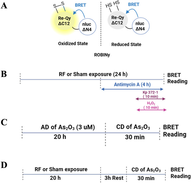
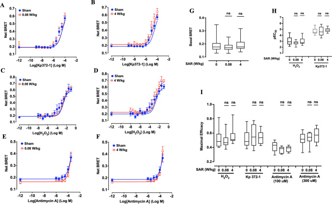
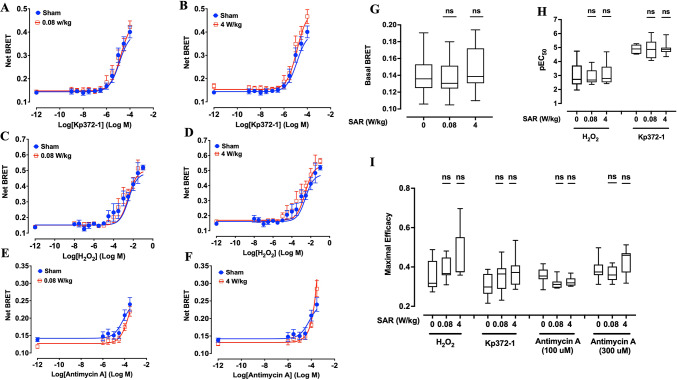
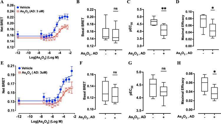
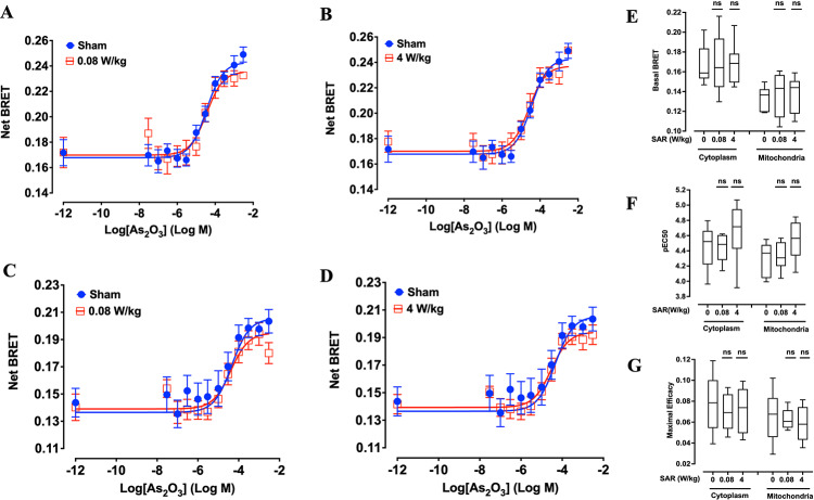
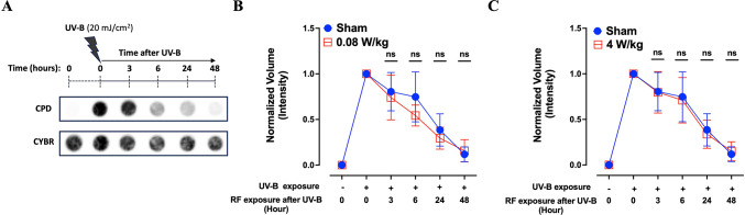
The rapid deployment of fifth-generation (5G) wireless networks has raised societal concerns regarding potential biological effects, particularly on human skin, due to the use of higher carrier frequencies that penetrate tissue less deeply. Consequently, whether 5G-modulated radiofrequency (RF) electromagnetic fields (EMFs) at 3.5 GHz affect oxidative stress and DNA repair in skin cells remains an open question. Using genetically encoded Bioluminescence Resonance Energy Transfer (BRET)-based biosensors targeted to the cytoplasm and mitochondria, we assessed whether exposure of human fibroblasts to 5G RF-EMF at specific absorption rates (SAR) of 0.08 and 4 W/kg for 24 h could alter basal reactive oxygen species (ROS) levels or potentiate the effects of known ROS inducers, including H₂O₂, Kp372-1, and Antimycin A. We also evaluated whether pre-exposure to 5G RF-EMF could induce an adaptive response (AR), by modulating ROS production following a subsequent challenge with arsenic trioxide (As₂O₃). Additionally, we investigated the impact of combined RF-EMF and ultraviolet-B (UV-B) exposure on the formation and repair of cyclobutane pyrimidine dimer (CPD) lesions in HaCaT keratinocytes. Our results showed no significant effect of 5G RF-EMF exposure, either alone or in combination with chemical ROS inducers, on oxidative stress markers in either compartment. Likewise, RF-EMF exposure did not induce an adaptive response to oxidative challenge, nor did it alter the kinetics or the efficiency of CPD repair by the nucleotide excision repair (NER) pathway. These findings support the conclusion that the exposure to 5G RF-EMF at 3.5 GHz up to 4 W/kg does not induce oxidative stress or impair DNA repair efficiency in human skin cells, within the experimental conditions tested.

Keywords: Bioluminescence resonance energy transfer (BRET); Cellular stress response; Cyclobutane pyrimidine dimer (CPD); DNA damage; Fifth generation (5G); Nucleotide excision repair (NER); Oxidative stress; Radiofrequency electromagnetic fields (RF-EMF); Reactive oxygen species (ROS).


Conflict of interest statement

Declarations. Competing interests: The authors declare no competing interests.

Figures

Fig. 1
Fig. 1
(A) Schematic representation of the BRET-based redox-sensitive biosensor ROBINy in both oxidized and reduced states (adapted from Fu et al.). ROBINy consists of nanoLuc ΔN4 (nluc lacking the N-terminal 4 amino acids) and Re-Qy ΔC12 (a redox-sensitive fluorescent acceptor lacking the C-terminal 12 amino acids). Under oxidative conditions, disulfide bonds form within Re-Qy, increasing its fluorescence and thus enhancing the energy transfer from nluc to Re-Qy, resulting in a higher BRET ratio. (BD) Timeline of experiments conducted in XP6BE human fibroblasts transfected with cytoplasmic or mitochondrial ROBINy. B) Direct ROS response assay: cells were sham- or RF-EMF-exposed (5G-modulated, 3.5 GHz) for 24 h. During the last 10 min of exposure, cells were treated with either H₂O₂ (0–100 mM) or Kp372-1 (0–100 µM). Alternatively, cells were treated with Antimycin A (0–300 µM) during the final 4 h of the exposure period. (C) Positive control for adaptive response: cells were pretreated with arsenic trioxide (As₂O₃, 3 µM, 20 h; adaptive dose, AD), then challenged with increasing concentrations (challenging dose, CD, 30 min). (D) RF-induced adaptive response assay: cells were exposed to RF-EMF or sham-exposed for 20 h, rested for 3 h, then challenged with CD of As₂O₃ for 30 min. BRET was measured immediately after the end of the RF exposure and/or chemical treatment.
Fig. 2
Fig. 2
Effect of 24-h exposure to 5G-modulated 3.5 GHz RF-EMF on ROS production induced by chemical stressors in XP6BE human fibroblasts transfected with the cytoplasmic ROBINy BRET probe. Cells were exposed for 24 h to sham or RF-EMF at 0.08 W/kg (A, C, E) or 4 W/kg (B, D, F), and challenged with increasing concentrations of Kp372-1 (A, B), H₂O₂ (C, D), or Antimycin A (E, F). Chemical stressors were applied during the final 10 min (for Kp372-1 and H₂O₂) or final 4 h (for Antimycin A) of RF exposure, as illustrated in Fig. 1B. BRET signals were recorded immediately after RF-EMF exposure, allowing real-time assessment of ROS dynamics. (GI) Box plots display quantitative analysis of basal BRET signal (G), pEC50 values of H2O2 and Kp372-1 (H), and maximal efficacy for each ROS inducer (I) across the different SAR conditions. Due to its limited potency, Antimycin A efficacy was calculated based only on responses to the two highest concentrations (100 and 300 µM). Statistical analysis was performed using Kruskal–Wallis test. No significant differences were observed between RF-exposed and sham groups (n = 8–9 per condition). n.s.: not significant.
Fig. 3
Fig. 3
Effect of 24-h exposure to 5G-modulated 3.5 GHz RF-EMF on ROS production induced by chemical stressors in XP6BE human fibroblasts transfected with the mitochondrial ROBINy BRET probe. Cells were exposed for 24 h to sham or RF-EMF at 0.08 W/kg (A, C, E) or 4 W/kg (B, D, F), and challenged with increasing concentrations of Kp372-1 (A, B), H₂O₂ (C, D), or Antimycin A (E, F). Chemical stressors were applied during the final 10 min (for Kp372-1 and H₂O₂) or final 4 h (for Antimycin A) of RF exposure, as described in Fig. 1B. BRET signals were acquired immediately after RF-EMF exposure, allowing real-time monitoring of mitochondrial ROS levels. (GI) Box plots summarize the quantitative effects of RF-EMF exposure on basal BRET signal (G), pEC50 values of H2O2 and Kp372-1 (H), and maximal efficacy for each ROS inducer (I) across the different SAR conditions. Due to its limited potency, Antimycin A efficacy was calculated based only on responses to the two highest concentrations (100 and 300 µM). Statistical analysis was performed using the Kruskal–Wallis and Mann–Whitney tests. No significant differences were observed between RF-exposed and sham groups (n = 8–9 depending on condition). n.s.: not significant.
Fig. 4
Fig. 4
Adaptive response to arsenic trioxide (As₂O₃) in XP6BE human fibroblasts expressing cytoplasmic or mitochondrial ROBINy BRET probes. Cells were pretreated with 3 µM As₂O₃ for 20 h to induce an adaptive dose (AD), followed by a 30-min challenging dose (CD) with increasing concentrations of As₂O₃, as shown in Fig. 1C. Panels A–D show the results obtained with the cytoplasmic BRET probe: dose–response curves (A), basal BRET signal (B), pEC₅₀ values of As2O3 (C), and maximal efficacy of As2O3 (D). Panels E–H show the corresponding data for the mitochondrial probe: dose–response curves (E), basal BRET signal (F), pEC₅₀ values of As2O3 (G), and maximal efficacy of As2O3 (H). A significant decrease in pEC₅₀ (panel C) and maximal efficacy (panels D and H) was observed in cells pretreated with the adaptive dose, demonstrating a measurable adaptive response to As₂O₃. Statistical analysis was performed using the Mann–Whitney test (*p < 0.05, **p < 0.01; n = 8–9 depending on experimental condition). n.s.: not significant.
Fig. 5
Fig. 5
Assessment of potential RF-EMF-induced adaptive response in XP6BE fibroblasts. Cells were exposed to sham or 5G-modulated 3.5 GHz RF-EMF at 0.08 or 4 W/kg for 20 h, followed by a 3 h resting phase. Then, increasing challenging dose (CD) concentrations of As₂O₃ were applied for 30 min before BRET measurement, as illustrated in Fig. 1D. (A and B) Dose response curves of As2O3 obtained with XP6BE fibroblast cells expressing the cytoplasmic ROBINy and being exposed to RF-EMF at SAR of 0.08 W/kg (A) or 4W/kg (B). (C and D) Dose response curves of As2O3 obtained with XP6BE fibroblast cells expressing the mitochondrial ROBINy BRET probe and being exposed to RF EMF at SAR of 0.08 W/kg (C) or 4 W/kg (D). (EG) Box plots showing basal BRET (E), pEC50 of As2O3 (F), and maximal efficacy of As2O3 (G) across the different SAR conditions. No adaptive response was observed following RF-EMF exposure, in contrast to As₂O₃-induced adaptation (Fig. 4). Statistical analysis was conducted using the Kruskal–Wallis Test. n = 8–9 depending on experimental conditions. n.s.: not significant.
Fig. 6
Fig. 6
Evaluation of DNA repair following UV-B and RF-EMF exposure in HaCaT human keratinocytes. (A) Dot-blot analysis of CPD (Cyclobutane Pyrimidine Dimers) at various time points (0–48 h) post UV-B exposure (20 mJ/cm2). (B, C) Quantification of CPD repair kinetics following UV-B exposure and subsequent RF-EMF exposure at 0.08 W/kg (B) or 4 W/kg (C). RF-EMF exposure did not significantly affect DNA repair kinetics. Statistical analysis was conducted using the Mann–Whitney Test between sham and RF-EMF exposed cells (n = 7). n.s.: not significant.

Similar articles

References

    1. ICNIRP. Guidelines for limiting exposure to electromagnetic fields (100 kHz to 300 GHz). Health Phys. 118, 483–524 (2020). - PubMed
    1. Baan, R. et al. Carcinogenicity of radiofrequency electromagnetic fields. Lancet Oncol.12, 624–626 (2011). - PubMed
    1. Gopal, B. G. A comparative study on 4G and 5G technology for wireless applications. IOSR J. Electron. Commun. Eng. IOSR-JECE10(6), 2278–2834 (2015).
    1. Mahmud, M. Cellular mobile technologies (1G to 5G) and massive MIMO. Int. J. Sci. Res. IJSR8, 929–937 (2019).
    1. De Iuliis, G. N., Newey, R. J., King, B. V. & Aitken, R. J. Mobile phone radiation induces reactive oxygen species production and DNA damage in human spermatozoa in vitro. PLoS ONE4, e6446 (2009). - PMC - PubMed

Substances

Grants and funding