. 2025 Aug 25;15(1):31301.
doi: 10.1038/s41598-025-14367-4.

Investigating the role of molecular coating in human corneal endothelial cell primary culture using artificial intelligence-driven image analysis

Affiliations

Investigating the role of molecular coating in human corneal endothelial cell primary culture using artificial intelligence-driven image analysis

Gauthier Travers et al. Sci Rep. .

Abstract

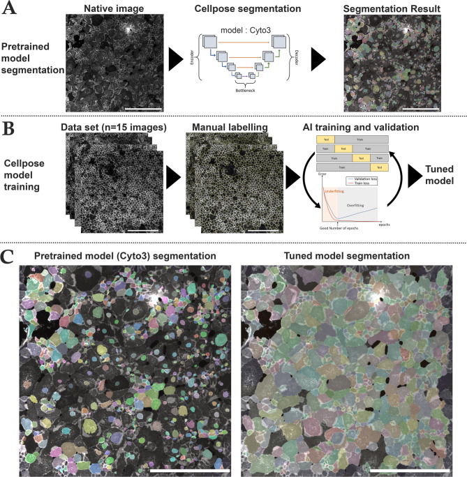
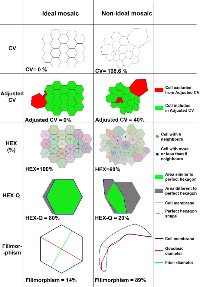
The monolayer of approximately 300,000 human corneal endothelial cells (hCECs) on the posterior surface of the cornea is essential to maintain transparency but is non-self-regenerative. Corneal blindness can currently only be treated by corneal transplantation, hindered by a global donor shortage, highlighting the need for developing tissue and/or cell therapy. The mass production of these advanced therapy medicinal products requires obtaining high-yield, high-quality endothelial cell cultures characterized by hexagonal shape, low size variability, and high endothelial cell density (ECD). Among the usual critical quality attributes which combine the expression of differentiation markers, ECD and cell morphological parameters, the latter are not optimally measured in vitro by conventional image analysis which poorly recognizes adherent cultured cells. We developed a high-performance automated segmentation using Cellpose algorithm and an original analysis method, improving the calculation of classical morphological parameters (coefficient of variation of cell area and hexagonality) and introducing new parameters specific to hCECs culture in vitro. Considering the importance of the extracellular matrix in vivo, and the panel of molecules available for coating cell culture plastics, we used these new tools to perform a comprehensive comparison of 13 molecules (laminins and collagens). We demonstrated their ability to discriminate subtle differences between cultures.

Keywords: Cell therapy; Endothelial cell density (ECD); High-performance automatic segmentation; Human corneal endothelial cells (hCECs); Immunohistochemistry; Molecular coating; Primary cell culture; Tissue-engineered endothelial keratoplasty (TEEK).


Conflict of interest statement

Declarations. Competing interests: The authors declare no competing interests.

Figures

Fig. 1
Fig. 1
Cell segmentation and AI training steps using Cellpose. (A) Steps for image segmentation with Cellpose which used an U-net model pre-trained on different cell types (Cyto3 model). The initial segmentation was not efficient on NCAM-labeled cultured corneal endothelial cells. Cell areas were coded by colors. (B) Steps for training a new model consisted in the selection of NCAM-labeled image of various quality (n = 15 images), followed by manual labeling and then a 4-fold cross validation training to optimize epochs and control segmentation quality of the newly trained model (tuned model). (C) Comparison of the same image segmented with the pre-trained Cyto3 model and with our tuned model. Scale bar = 500 μm on each image.
Fig. 2
Fig. 2
Parameters for quantifying hCECs morphology comparing ideal mosaic and non-ideal mosaic. CV Coefficient of variation of cell area, HEX percentage of hexagonal cells. Details for each parameter and their calculation method can be found in the supplementary methods.
Fig. 3
Fig. 3
Validation of the AI-based method for quantifying the various hCECs morphological parameters. (A) Endothelial cell cultures of highly variable quality used for AI algorithm validation. Cell lateral membranes were labeled with anti-NCAM (in green) and the nuclei were counterstained using DAPI (in blue). The images were taken in the center of each well at x10 magnification with a zoom x6 in the center of the image. Image scale bar = 300 μm, zoom scale bar = 50 μm. (B) Evaluation of 6 parameters across 4 different culture qualities. Graph illustrating 6 objective evaluation parameters across 4 culture qualities: (1) EndoMT cells at passage 7; (2) Mixed population quality at passage 5; (3) High quality – young donor (below 30 yo) at passage 4; (4) High quality – aged donor (over 30 yo), at passage 11. ns = non-significant = p-value > 0.05, *p-value < 0.05, **p-value < 0.01, ***p-value < 0.001.
Fig. 4
Fig. 4
Endothelial quality score (EQS) for evaluating cell culture quality based on morphological and ECD criteria. EQS was calculated from the z-score of each of the 6 chosen parameters (ECD, Hexagonality, Quality of Hexagonality, CV, Adjusted CV, Filimorphism). Graph illustrating 6 objective evaluation parameters across 4 culture qualities: (1) EndoMT cells at passage 7; (2) Mixed population quality at passage 5; (3) High quality – young donor (below 30 yo) at passage 4; (4) High quality – aged donor (over 30 yo), at passage 11. *=p-value < 0.05, **=p-value < 0.01, ***=p-value < 0.001).
Fig. 5
Fig. 5
Representative example of the effects of coating molecules on hCECs cultured in vitro. Molecular names were expressed on the top left of each image. Images were taken in the center of each well at x40 magnification. The control represented a well without coating. Cell lateral membranes were stained with anti-NCAM (green) and the nuclei were counterstained using DAPI (blue). The donor used for this example was “c” in Table 1. Scale bars are placed in the bottom-right images of columns IV and CTRL (100 μm).
Fig. 6
Fig. 6
Summary of the mixed model analysis for coating molecules. (A) The table presented the ratio, p-value, and confidence interval, highlighting significant differences between the tested coating molecules and the control. The color scale indicates the level of significance, with darker green representing higher significance and lighter green representing lower significance. (B) Forest plot of molecule expression ratios according to the evaluated criterion. This forest plot illustrated the expression ratios of different coating molecules based on the evaluated criterion. Each point represents the mean ratio value, while the vertical bars indicate the corresponding confidence intervals (lower and upper bounds). The vertical line at y = 1 served as the reference value compared to the uncoated control. A ratio greater than 1 suggested superiority compared to the control, whereas a ratio lower than 1 indicated inferiority compared to the control. If the ratio of the parameters ECD, Hexagonality, and HEX-Q is greater than that of the control, the culture quality is better (green zone); otherwise, it is worse (red zone). If the ratio of the parameters CV, Adjusted CV, and Filimorphism is greater than that of the control, the culture quality is worse (red zone); otherwise, it is better (green zone). (C) Forest plot of molecule expression ratios (compared to the uncoated control) according to the endothelial quality score (EQS) (including all 6 evaluations parameters). Each point represented the mean ratio value, while the horizontal bars indicated the corresponding confidence intervals (lower and upper bounds).

Similar articles

References

    1. Liu, C., Gao, Z. Q., Li, J. & Zhou, Q. et « Identification of novel therapeutic targets for Fuchs’ endothelial corneal dystrophy based on gene bioinformatics analysis », PLOS ONE, vol. 17, no 3, p. e0264018, mars (2022). 10.1371/journal.pone.0264018 - PMC - PubMed
    1. Chen, X., Xu, J. & Chen, X. Yao, « cataract: advances in surgery and whether surgery remains the only treatment in future ». Adv. Ophthalmol. Pract. Res.1, 100008. 10.1016/j.aopr.2021.100008 (nov. 2021). no 1. - PMC - PubMed
    1. Gain, P. et al. « Global Survey of Corneal Transplantation and Eye Banking », JAMA Ophthalmol, vol. 134, no 2, pp. 167–173, févr. (2016). 10.1001/jamaophthalmol.2015.4776 - PubMed
    1. Crouzet, E. et al. « Tissue engineered endothelial keratoplasty in rabbit: tips and tricks », Acta Ophthalmologica, vol. 100, no 6, pp. 690–699, (2022). 10.1111/aos.15081 - PubMed
    1. Parekh, M. et al. « Biomaterials for corneal endothelial cell culture and tissue engineering », J Tissue Eng, vol. 12, p. 2041731421990536, janv. (2021). 10.1177/2041731421990536 - PMC - PubMed

LinkOut - more resources