. 2025 Aug 25;15(1):31220.
doi: 10.1038/s41598-025-17065-3.

Cholecystokinin-expressing GABA neurons elicit long-term potentiation in the cortical inhibitory synapses and attenuate sound-shock associative memory

Affiliations

Cholecystokinin-expressing GABA neurons elicit long-term potentiation in the cortical inhibitory synapses and attenuate sound-shock associative memory

Ge Zhang et al. Sci Rep. .

Abstract

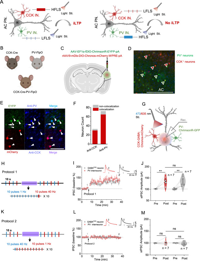
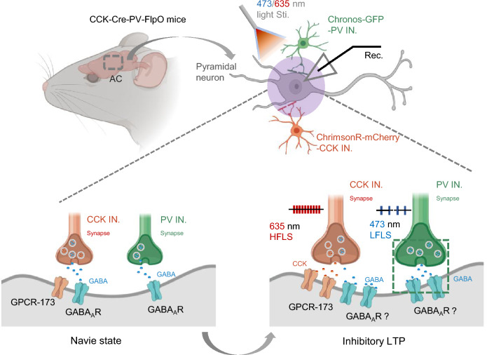
Neuronal interactions between inhibitory and excitatory neurons play a pivotal role in regulating the balance of excitation and inhibition in the central nervous system (CNS). Consequently, the efficacy of inhibitory/excitatory synapses profoundly affects neural network processing and overall neuronal functions. Here, we describe a novel form of long-term potentiation (LTP) induced at cortical inhibitory synapses and its behavioral consequences. We show that high-frequency laser stimulation (HFLS) of GABAergic neurons elicit inhibitory LTP (i-LTP) in pyramidal neurons of the auditory cortex (AC). The selective activation of cholecystokinin-expressing GABA (GABACCK) neurons is essential for the formation of HFLS-induced i-LTP, rather than the classical parvalbumin (PV) neurons and somatostatin (SST) neurons. Intriguingly, i-LTP can be evoked in the AC by adding the exogenous neuropeptide CCK when PV neurons and SST neurons are selectively activated in PV-Cre and SST-Cre mice, respectively. Additionally, we discovered that low-frequency laser stimulation (LFLS) of PV neurons paired with HFLS of GABACCK neurons potentiates the inhibitory effect of PV interneurons on pyramidal neurons, thereby generating heterosynaptic i-LTP in the AC. Notably, light activation of GABACCK neurons in CCK-Cre mice significantly attenuates sound- shock associative memory, while stimulation of PV neurons does not affect this memory in PV-Cre mice. In conclusion, these results demonstrate a critical mechanism regulating the excitation-inhibition balance and modulating learning and memory in cortical circuits. This mechanism might serve as a potential target for the treatment of neurological disorders, including epilepsy and Alzheimer's disease.

Keywords: Auditory cortex; Cholecystokinin; Interneuron; Long-term potentiation; Neurological disorders.


Conflict of interest statement

Declarations. Competing interests: The authors declare no competing interests.

Figures

Fig. 1
Fig. 1
HFLS of GABA neuron induced i-LTP on pyramidal neuron in the AC. (A) Schematic demonstration of how GABA neurons exhibit its neuronal effects on pyramidal neurons in auditory cortex (AC PN.). (B) Schematic illustration of Cre-dependent AAV vector (DIO-Chronos-mCherry) injection into the AC of Vgat-Cre mice to specifically infect GABA neurons. (C) Schematic diagram of virus injection into the AC of Vgat-Cre mice (AAV9-mDlx-DIO-Chronos-mCherry-WPRE-pA, 6.15E + 12 vg/mL, 200 nL for two different sites). (D) Confocal image of virus expression in the AC. White arrows indicate the colocalization of antibody-GAD67 with mCheryy expressed GABA positive neurons. Scale bar: 50 µm. (E) Schematic depiction of the IPSC recording of the GABA neuron in the AC (left); A representative trace of the regular spiking of a pyramidal neuron (right). (F) Protocols (HFLS and LFLS) for stimulating the Chronos expressed GABA neuron during the IPSC recording. (G) Normalized amplitude of IPSC before and after 473 nm wavelength LFLS (grey) and HFLS (red). (H) Statistical comparison of amplitudes of IPSC before and after 473 nm wavelength LFLS (grey) and HFLS (red). (I) Quantitative analyses of sIPSC frequency before and after the 473 nm wavelength LFLS. (J) Quantitative analyses of sIPSC amplitude before and after the 473 nm wavelength LFLS. (K) Quantitative analyses of sIPSC frequency before and after the 473 nm wavelength HFLS. (L) Quantitative analyses of sIPSC amplitude before and after the 473 nm wavelength HFLS. *p < 0.05, **p < 0.01, ***p < 0.001; ns, not significant. Data are reported as mean ± SEM.
Fig. 2
Fig. 2
CCK-GABA neurons are crucial for HFLS-induced i-LTP. (A) Schematic demonstration of which type of interneuron contribute to the iLTP formation in pyramidal neurons of auditory cortex (AC PN.). (B) Schematic illustration of Cre-dependent AAV vector (DIO-Chronos-mCherry; 6.15E + 12 vg/mL, 200 nL for two different sites) injection into the AC of CCK-Cre mice and PV-Cre mice to specifically infect GABAergic neurons, respectively. (C) Confocal image of virus expression in the AC of CCK-Cre mice (upper) and PV-Cre mice (bottom). White arrows indicate the colocalization of antibody-CCK/PV with mCheryy expressed CCK/PV positive neurons. Scale bar: 50 µm. (D) Schematic depiction of the IPSC recording of GABACCK/PV interneuron in the AC. (E) Normalized amplitude of IPSC before and after 473 nm wavelength HFLS in CCK-Cre mice (red) and PV-Cre mice (grey), respectively. (F) Statistical comparison of amplitude of IPSC before and after 635 nm wavelength HFLS in CCK-Cre mice (red) and PV-Cre mice (grey), respectively. (G) Representative sIPSC traces of GABACCK neurons before (upper) and after (bottom) HFLS in the CCK-Cre mice. (H) Representative sIPSC traces of PV interneurons before (upper) and after (bottom) HFLS in the PV-Cre mice. (I) Quantitative analyses of sIPSC frequency before and after the 473 nm wavelength HFLS in CCK-Cre mice (red) and PV-Cre mice (grey), respectively. (J) Quantitative analyses of sIPSC amplitude before and after the 473 nm wavelength LFLS in CCK-Cre mice (red) and PV-Cre mice (grey), respectively. *p < 0.05, **p < 0.01, ***p < 0.001; ns, not significant. Data are reported as mean ± SEM.
Fig. 3
Fig. 3
Exogenous CCK facilitates the formation of i-LTP in the AC. (A) Schematic demonstration of whether exogenous CCK interacts with PV interneuron to elicit iLTP in pyramidal neurons of auditory cortex (AC PN.). (B) Schematic depiction of the IPSC recording of PV interneuron in the AC of PV-Cre mice. (C) Normalized amplitude of IPSC before and after perfusion with CCK-8s in HFLS group (red) and LFLS group (grey), respectively. (D) Statistical comparison of the amplitude of IPSC before and after perfusion with CCK-8s in both groups. (E) Schematic demonstration of whether exogenous CCK interacts with SST interneuron to elicit iLTP in pyramidal neurons of auditory cortex (AC PN.). (F) Schematic depiction of the IPSC recording of the SST interneuron in the AC of the SST-Cre mcie. (G) Normalized amplitude of IPSC before and after perfusion with CCK-8s in HFLS group (red) and LFLS group (grey), respectively. (H) Statistical comparison of the amplitude of IPSC before and after perfusion with CCK-8s in both groups. *p < 0.05, **p < 0.01, ***p < 0.001; ns, not significant. Data are reported as mean ± SEM.
Fig. 4
Fig. 4
Potentiation of inhibition effects of PV-interneuron on pyramidal neuron requires high-frequency activation of GABACCK neuron. (A) Schematic demonstration of whether HFLS of CCK interneurons or PV interneurons facilitate the iLTP formation in AC PN. (B) Schematic illustration of the acquisition of CCK-Cre-PV-FlpO pups by crossing CCK-Cre mice with PV-Flop mice. (C) Mixture of AAV9-mDlx-DIO-Chronos-mCherry-WPRE-pA and AAV9-fDIO-ChrmosonR-EYFP-WPRE-pA was injected into the AC area of CCK-Cre-PV-FlpO mice (DIO-Chronos-mCherry/-fDIO-ChrimsonR-EYFP-pA; 6.15E + 12 vg/mL, 200 nL for two different sites). (D) Fluorescent image showing AAV expression in AC area of CCK-Cre-PV-FlpO mice. (E) Confocal image of two kinds of AAV expression in pyramidal neurons in AC area. White arrows indicate the colocalization of antibody-CCK/antibody-PV with mCheryy expressed CCK and PV positive neurons, respectively. Scale bar: 50 µm. (F) Schematic depiction of two kinds of lights to separately evoke the IPSC in Chronos expressed GABACCK neuron and ChrimsonR infected PV neuron in the AC area of CCK-Cre-PV-FlpO mice. (G) Protocol consist of 473 nm HFLS and 635 nm LFLS for activating the Chronos expressed GABACCK neuron and ChrimsonR infected PV neuron, respectively. (H) Normalized amplitude of IPSC in GABACCK neuron and PV neuron before and manipulating the protocol 1, respectively. (I) Statistical comparison of the amplitude of IPSCs before and after manipulating the protocol 1 in both groups. (J) Protocol consist of 473 nm LFLS and 635 nm HFLS for activating the Chronos expressed GABACCK neuron and ChrimsonR infected PV neuron, respectively. (K) Normalized amplitude of IPSC in GABACCK neuron and PV neuron before and manipulating the protocol 2, respectively. (L) Statistical comparison of the amplitudes of IPSCs before and after manipulating the protocol 2 in both groups. *p < 0.05, **p < 0.01, ***p < 0.001; ns, not significant. Data are reported as mean ± SEM.
Fig. 5
Fig. 5
Potentiation of inhibition effects of PV-interneuron on pyramidal neuron requires high-frequency activation of GABACCK neuron. (A) Schematic demonstration of whether HFLS of CCK interneurons or PV interneurons affects animals’ fear memory. (B) Schematic depiction of behavioral diagram of sound-shock association memory. (C) The drawing of the protocol of the behavioral diagram. (D) Schematic illustration of AAV injection and fiber implantation in AC of experimental mice. (E) Fluorescent image showing the AAV expression and fiber track in AC area. (F) Statistical analysis of the freezing proportion in three different groups of mice. *p < 0.05, **p < 0.01, ***p < 0.001; ns, not significant. Data are reported as mean ± SEM.
Fig. 6
Fig. 6
Schematic illustration depicts a possible cellular mechanism that HFLS of GABACCK release via by both postsynaptic GPCR173 receptor and GABAA receptors produces LTP and impairs sound-shock associative memory. HFLS of Chronos expressed GABACCK neuron pairs with LFLS of ChrimsonR infected PV neuron triggers CCK released from the presynaptic terminals that facilitates the potentiates the inhibitory effects of both interneurons on post-synapses of pyramidal neurons in the AC area.

Similar articles

References

    1. Xu, Z., Geron, E., Pérez-Cuesta, L. M., Bai, Y. & Gan, W. B. Generalized extinction of fear memory depends on co-allocation of synaptic plasticity in dendrites. Nat. Commun.14(1), 503. 10.1038/s41467-023-35805-9 (2023). - PMC - PubMed
    1. Jeong, Y. et al. Synaptic plasticity-dependent competition rule influences memory formation. Nat. Commun.12(1), 3915. 10.1038/s41467-021-24269-4 (2021). - PMC - PubMed
    1. Caya-Bissonnette, L. & Béïque, J. C. Half a century legacy of long-term potentiation. Curr. Biol.34(13), R640–R662. 10.1016/j.cub.2024.05.008 (2024). - PubMed
    1. Scott, H. L. et al. Evidence for long-term potentiation in phospholipid membranes. Proc. Natl. Acad. Sci.119(50), e2212195119. 10.1073/pnas.2212195119 (2022). - PMC - PubMed
    1. Bliss, T. V. P. & Collingridge, G. L. A synaptic model of memory: Long-term potentiation in the hippocampus. Nature361(6407), 31–39. 10.1038/361031a0 (1993). - PubMed

LinkOut - more resources