. 2025 Oct;39(10):6943-6954.
doi: 10.1007/s00464-025-12081-w. Epub 2025 Aug 25.

Back-table specimen scanning using gantry-free hybrid hSPECT/LiDAR imaging: a feasibility study during PSMA-radioguided surgery

Affiliations

Back-table specimen scanning using gantry-free hybrid hSPECT/LiDAR imaging: a feasibility study during PSMA-radioguided surgery

Giusi Pisano et al. Surg Endosc. 2025 Oct.

Abstract

Introduction: Prostate-specific membrane antigen (PSMA) targeted precision surgery is becoming increasingly popular. However, the relatively low levels of PSMA-receptor expression and background signal can hinder in vivo lesion detection and margin evaluation. Back-table imaging (ex vivo) potentially provides a means to confirm surgical accuracy. For 99mTc-PSMA-radioguided surgery, an innovative gantry-free hybrid imaging technique has recently been proposed, namely handheld single-photon emission computed tomography (hSPECT) combined with light detection and ranging (LiDAR). This study aimed to assess the feasibility and performance of hSPECT/LiDAR in analyzing tissue specimens excised after robotic 99mTc-PSMA-radioguided surgery.

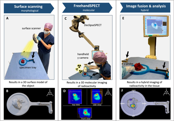
Methods: We included samples from 5 prostate cancer patients undergoing primary or salvage robot-assisted resection of 99mTc-PSMA-I&S avid lesions that were identified using a drop-in gamma probe. 12 samples (1 prostatic tissue, 1 local recurrence tissue, 10 lymph nodes) were analyzed ex vivo using a custom-built specimen tray, including an optical reference tracker for scan registration. LiDAR was used to acquire a surface scan of the specimens, and the 3D OBJ image output was fused with the 3D DICOM of a hSPECT obtained using a handheld gamma camera and DeclipseSPECT tracking system.

Results: hSPECT/LiDAR imaging provided accurate representation of the 99mTc-PSMA-I&S uptake within the specimens. In 8 samples, it helped to confirm a true positive lesion. In the remaining 4 samples, non-visualization aligned with negative histopathology (true negative). A strong correlation was found between PSMA-hSPECT/LiDAR and PSMA-PET/CT (p < 0.05), but no correlation could be established with PSMA-SPECT/CT (p = 0.515). The count rates fount in the scan correlated to tumor size (p = 0.016) and were not influenced by the overall specimen's size (p = 0.558).

Conclusion: We present the technical feasibility of a new 3D hybrid modality (hSPECT/LiDAR) that allows back-table assessment of surgical specimens from the already well validated robotic 99mTc-PSMA-radioguided surgery workflow.

Keywords: Image-guided surgery; PSMA SPECT/CT/LiDAR; Prostate cancer; Radioguided surgery; Specimen scanning; Surface scanning.


Conflict of interest statement

Declarations. Disclosures: BAÇ is an employee of Crystal Photonics; however, the company had no role in the design or reporting of the study. GP, MNvO, VAO, ACB, LJS, DDDR, HGvdP, PJvL, FWBvL have no disclosures to state.

Figures

Fig. 1
Fig. 1
Clinical workflow of this study starts with patients undergoing a diagnostic PSMA-PET/CT weeks before the procedure, for staging or follow-up purpose. If selected for robot-assisted radioguided surgery, patients underwent 99mTc-PSMA-I&S intravenous injection the day before surgery, and SPECT/CT acquisition on the morning of surgery. Intraoperatively, radioactivity was detected through the drop-in probe during robot-assisted surgery. After removal of the specimens, the ex vivo counts were measured using a handheld gamma probe and subsequently examined by a specimen scan before being sent to pathology for final analysis
Fig. 2
Fig. 2
The setup and instrumentations of the specimen scanning are depicted in sequence, in the case of a primary prostate specimen. First of all, surface scanning of the tissue (Artec Eva) was performed (black arrows indicate optical reference tracker) (A) to obtain a 3D surface model of the excised tissue specimen (B). Gantry-free SPECT was acquired using a declipseSPECT system combined with an optically tracked CrystalCam (C) to generate the images of 99mTc-PSMA distribution (D). Lastly, the LiDAR scan and the molecular 99mTc-PSMA hSPECT were fused and visualized, enabling a real-time examination of the tissue from different angles employing a handheld gamma probe as pointer (E). As a final result, a hybrid imaging of the radioactivity distribution within the tissue surface was generated (F)
Fig. 3
Fig. 3
Case 3 from Table 1–Displaying a case of recurrent cancer, of a patient who underwent RALP and pelvic lymph node dissection in 2017 for a GS 7 (3 + 4) acinar adenocarcinoma with no additional therapies. At that time, pathology resulted in a pT2aN0Mx, R0. At follow-up PSMA-PET/CT imaging, a PSMA-avid node was seen in the left pelvis (A, red arrow), not intense on SPECT/CT (B). At hSPECT/LiDAR specimen scanning, radiotracer uptake was visible inside the tissue (C, red arrow). At histopathological analysis (D, H&E left 1 mm magnification, right 200 μm magnification), the specimen showed to contain thick nerve bundles surrounded by acinar type adenocarcinoma, with adenocarcinoma localized also in the fibrous tissue. Again, the positive histology correlated with our images
Fig. 4
Fig. 4
A Radar chart of the primary prostate cancer case (Case 1 from Table 1) depicting radioactivity localization by hSPECT/LiDAR specimen scanning, PSMA-PET/CT, PSMA-SPECT/CT. Each axis represents a different variable: prostate zones (base, midgland, apex), focality (unifocal vs. multifocal), and involvement of seminal vesicles. The radial scale (from 0 to 5) represents the score of radioactivity localization for the modalities. A score of 0 suggests no radioactivity (negative), and a score of 5 indicates strong radioactivity (positive). The same parameters were correlated with tumor presence at histopathology (0: negative, 5: positive). B and C Scatter plot illustrating the relationship between respective specimen size (mm, x-axis) or tumor size (mm, x-axis) with count rate (counts/s, y-axis). Each data point represents an individual observation. A linear regression trend line (red) has been applied to visualize the overall pattern
Fig. 5
Fig. 5
Case 4 from Table 1- In this recurrence case, two nodes were scanned with hSPECT/LiDAR after robot-assisted radioguided surgery, one pararectal LN (suspicious) and one internal iliac LN (control). At 3D Slicer visualization of radioactivity A, two spots of signal could be seen at axial and coronal views (white and yellow arrows), that could have been attributed to the two LNs. At specimen scanning hybrid display B, the highest radioactivity spot clearly felt into the pararectal LN (yellow arrow), while the other spot being possibly attributable to a contamination on the specimen tray (white arrow)

References

    1. Donin NM, Reiter RE (2018) Why targeting PSMA is a game changer in the management of prostate cancer. J Nucl Med 59:177–182 - PMC - PubMed
    1. Maurer T, Gschwend JE, Rauscher I, Souvatzoglou M, Haller B, Weirich G et al (2016) Diagnostic efficacy of (68)gallium-PSMA positron emission tomography compared to conventional imaging for lymph node staging of 130 consecutive patients with intermediate to high risk prostate cancer. J Urol 195:1436–1443 - PubMed
    1. Perera M, Papa N, Roberts M, Williams M, Udovicich C, Vela I et al (2020) Gallium-68 prostate-specific membrane antigen positron emission tomography in advanced prostate cancer-updated diagnostic utility, sensitivity, specificity, and distribution of prostate-specific membrane antigen-avid lesions: a systematic review and meta-analysis. Eur Urol 77:403–417 - PubMed
    1. Hofman MS, Lawrentschuk N, Francis RJ, Tang C, Vela I, Thomas P et al (2020) Prostate-specific membrane antigen PET-CT in patients with high-risk prostate cancer before curative-intent surgery or radiotherapy (proPSMA): a prospective, randomised, multicentre study. Lancet 395:1208–1216 - PubMed
    1. Schottelius M, Wirtz M, Eiber M, Maurer T, Wester H-J (2015) [(111)In]PSMA-I&T: expanding the spectrum of PSMA-I&T applications towards SPECT and radioguided surgery. EJNMMI Res 5:68 - PMC - PubMed

MeSH terms

LinkOut - more resources