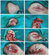
Potential of tadalafil and tadalafil-cellulose nanocomposite in preventing postsurgical abdominal adhesions in a rat cecal abrasion model
- PMID: 40855170
- PMCID: PMC12378406
- DOI: 10.1038/s41598-025-14894-0
Potential of tadalafil and tadalafil-cellulose nanocomposite in preventing postsurgical abdominal adhesions in a rat cecal abrasion model
Abstract
The formation of postoperative intra-abdominal adhesions is a significant challenge in veterinary practice worldwide. Thus, several attempts have been made to identify agents that prevent the occurrence of these postsurgical adhesions. However, finding an ideal and effective agent remains a challenge. Herein, we investigate the potential of tadalafil and tadalafil/cellulose composite as promising therapeutics for preventing postsurgical intra-abdominal adhesions. A cecal abrasion model was established in 30 rats, which either left untreated or treated with tadalafil, cellulose, or tadalafil/cellulose. After 2 weeks, the adhesion formation was evaluated based on gross appearance, oxidative stress markers, pro-inflammatory cytokines, histopathological analysis, and immunohistochemical staining. Compared to the adhesion group, gross and histopathological findings revealed that both the tadalafil and cellulose groups significantly decreased adhesion formation, with better results observed after tadalafil treatment. Importantly the tadalafil/cellulose treatment completely prevented adhesion formation. Additionally, the treated groups showed reduced levels of malondialdehyde (MDA), tumor necrosis factor-alpha (TNF-α), and interleukin-6 (IL-6), while increasing the level of reduced glutathione (GSH) compared to the adhesion group. Furthermore, the treated groups reduced the expression of macrophage markers. These findings suggest that the intra-abdominal application of tadalafil and tadalafil/cellulose following abdominal surgery holds promise as a clinical strategy to prevent postsurgical intra-abdominal adhesions, with tadalafil/cellulose demonstrating superior efficacy.
Keywords: Adhesions; Anti-adhesion; Cellulose; Intra-abdominal; Postsurgical; Tadalafil.
© 2025. The Author(s).
Conflict of interest statement
Declarations. Competing interests: The authors declare no competing interests.
Figures


Similar articles
-
Barrier agents for adhesion prevention after gynaecological surgery.Cochrane Database Syst Rev. 2015 Apr 30;2015(4):CD000475. doi: 10.1002/14651858.CD000475.pub3. Cochrane Database Syst Rev. 2015. Update in: Cochrane Database Syst Rev. 2020 Mar 22;3:CD000475. doi: 10.1002/14651858.CD000475.pub4. PMID: 25924805 Free PMC article. Updated.
-
Comparison of simvastatin gel and amnion membrane gel for preventing postoperative intra-abdominal adhesion formation in rat model.Injury. 2025 Aug;56(8):112541. doi: 10.1016/j.injury.2025.112541. Epub 2025 Jun 22. Injury. 2025. PMID: 40582291
-
Systemic pharmacological treatments for chronic plaque psoriasis: a network meta-analysis.Cochrane Database Syst Rev. 2021 Apr 19;4(4):CD011535. doi: 10.1002/14651858.CD011535.pub4. Cochrane Database Syst Rev. 2021. Update in: Cochrane Database Syst Rev. 2022 May 23;5:CD011535. doi: 10.1002/14651858.CD011535.pub5. PMID: 33871055 Free PMC article. Updated.
-
Systemic pharmacological treatments for chronic plaque psoriasis: a network meta-analysis.Cochrane Database Syst Rev. 2017 Dec 22;12(12):CD011535. doi: 10.1002/14651858.CD011535.pub2. Cochrane Database Syst Rev. 2017. Update in: Cochrane Database Syst Rev. 2020 Jan 9;1:CD011535. doi: 10.1002/14651858.CD011535.pub3. PMID: 29271481 Free PMC article. Updated.
-
Intra-peritoneal prophylactic agents for preventing adhesions and adhesive intestinal obstruction after non-gynaecological abdominal surgery.Cochrane Database Syst Rev. 2009 Jan 21;(1):CD005080. doi: 10.1002/14651858.CD005080.pub2. Cochrane Database Syst Rev. 2009. PMID: 19160246
References
-
- Salciccia, A. et al. Surgical closure of equine abdomen, prevention, and management of incisional complications. J. Vis. Exp.10.3791/65546 (2024). - PubMed
-
- Nichols, S. & Fecteau, G. Surgical management of abomasal and small intestinal disease. Vet. Clin. Food Anim. Pract.34, 55–81. 10.1016/j.cvfa.2017.10.007 (2018). - PubMed
-
- Fossum, T. W. in Small Animal Surgery (ed Terry W. Fossum) Ch. Soft Tissue Surgery, 512–539 (2019).
MeSH terms
Substances
LinkOut - more resources
Full Text Sources
Medical

