. 2025 Aug 25;22(1):24.
doi: 10.1186/s12983-025-00575-1.

Structure and function of the ovipositor of the encyrtid wasp Microterys flavus

Affiliations

Structure and function of the ovipositor of the encyrtid wasp Microterys flavus

Robin Kraft et al. Front Zool. .

Abstract

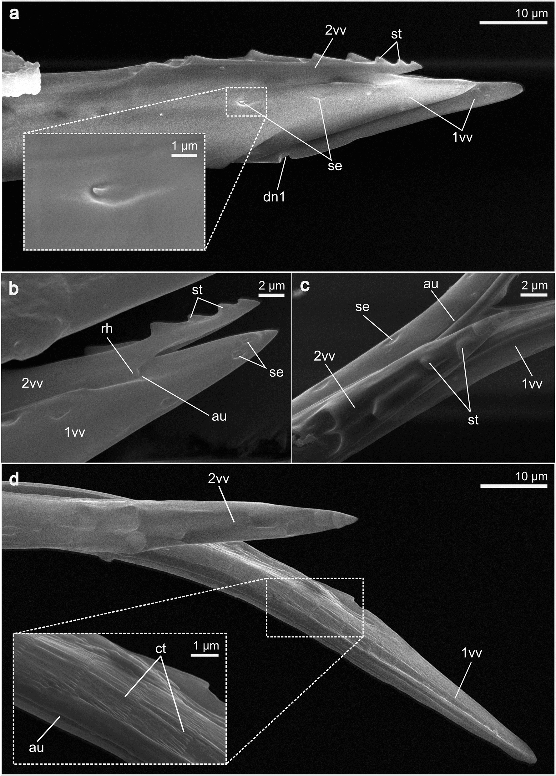
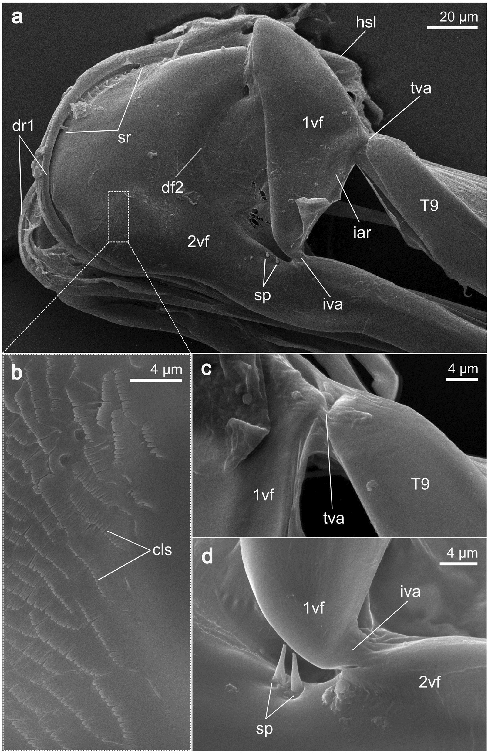
Background: Oviposition is crucial for the reproductive success of parasitoid insects and, hence, ovipositor structure and oviposition behaviour have probably played a central role in their adaptive evolution. However, various mechanical and functional aspects of the musculoskeletal ovipositor system are still not fully understood, especially within the enormously diverse parasitoid wasps, e.g. the minute and understudied Encyrtidae (Chalcidoidea). Some encyrtid wasps are specialized in parasitising insect plant pests and thus play an important ecological and economic role. We have examined all inherent cuticular elements and muscles of the ovipositor of the encyrtid wasp Microterys flavus to improve our understanding of its mechanics and mode of function. We provide a detailed 3D model based on a synchrotron X-ray phase-contrast microtomography (SR-µCT) dataset and have analysed microstructures on the cuticular ovipositor elements by using scanning electron microscopy (SEM). We have also conducted an in vivo documentation of the oviposition process of female M. flavus wasps on their host, the scale insect Coccus hesperidum.

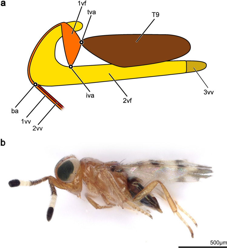
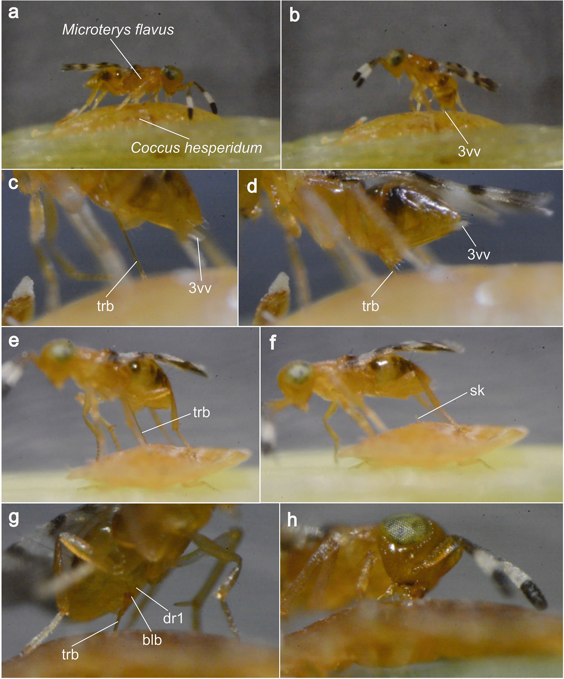
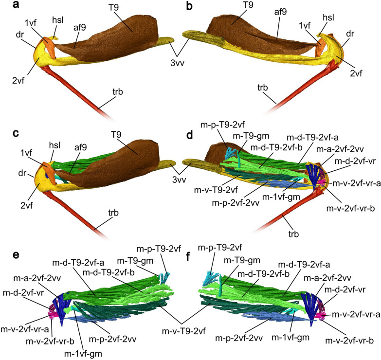
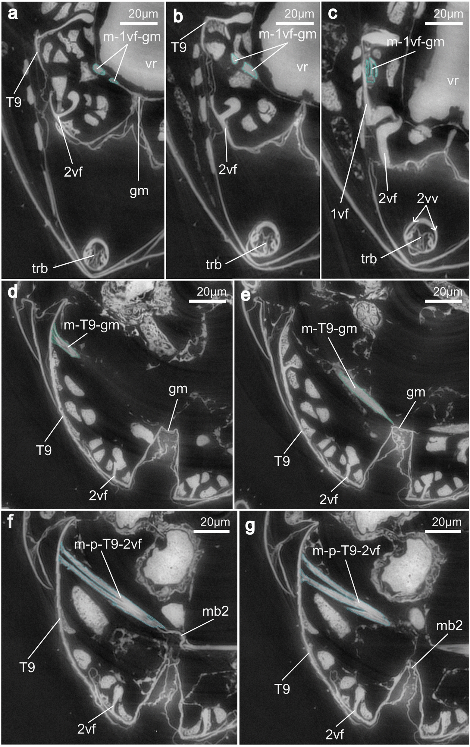
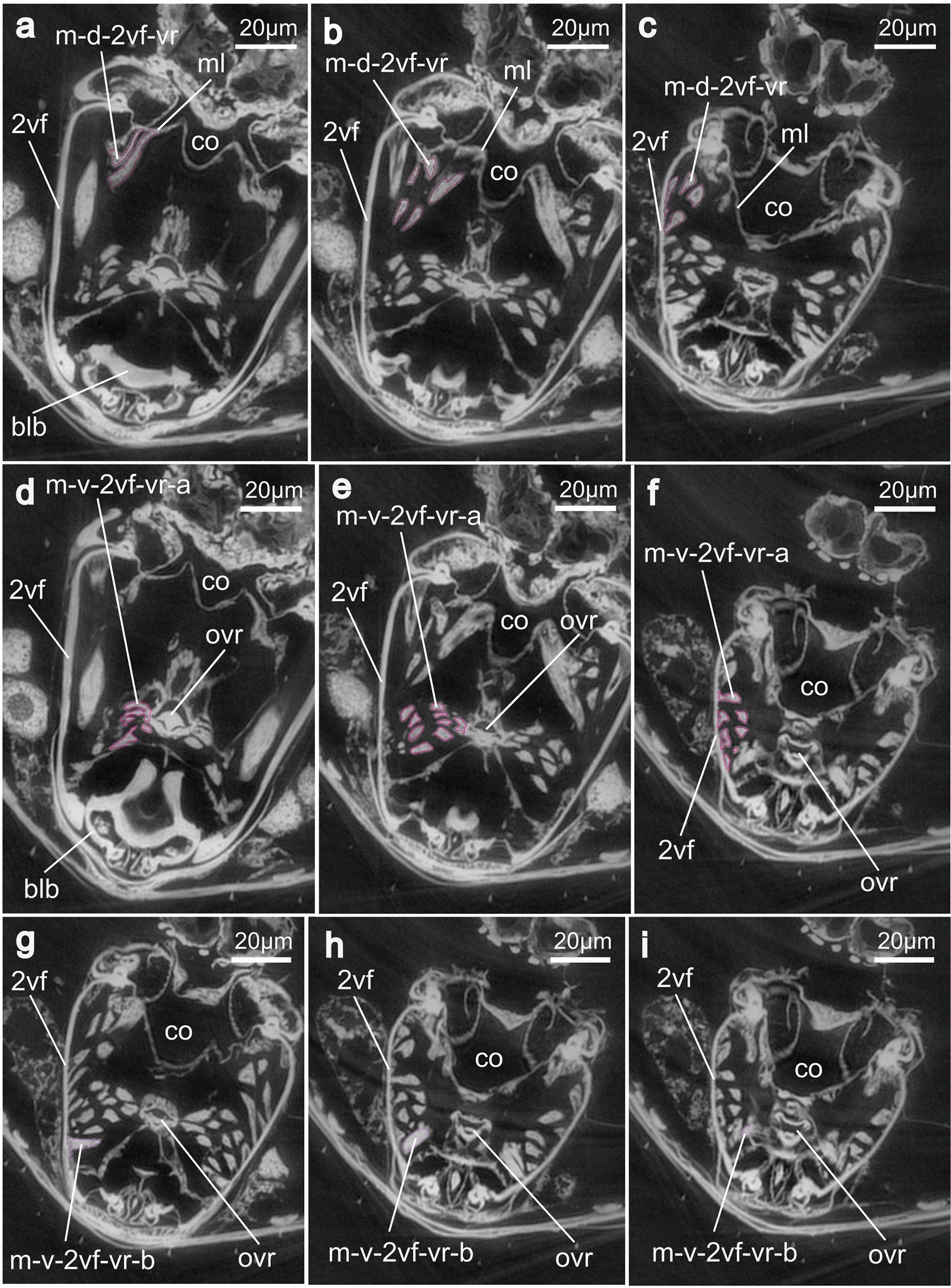
Results: Based on morphological analyses, we have identified all elements of the musculoskeletal ovipositor system in M. flavus, consisting of two pairs of valvifers, three pairs of valvulae, the female T9 (9th abdominal tergum), and a set of nine paired ovipositor muscles. Three of these muscles (1st valvifer-genital membrane muscle, ventral 2nd valvifer-venom gland reservoir muscle, T9-genital membrane muscle) have only recently been discovered in pteromalid wasps but have not yet been described for encyrtids. Our behavioural analysis of the motion patterns during the various phases of parasitization has elucidated the oviposition process, which consists of penetration of the host's body, assessment of the host's internal organs, envenomation, egg deposition, and potential host feeding.

Conclusions: Based on our studies of the structure of the ovipositor system of the encyrtid wasp M. flavus, we have developed a functional model of the underlying working mechanism of all ovipositor movements observed during the oviposition process, thereby improving our understanding of a possible key trait contributing to the evolutionary success of a highly diverse group of chalcidoid wasps.

Keywords: Chalcidoidea; Encyrtidae; Functional morphology; Hymenoptera; Ovipositor; Parasitoid; Terebra.


Conflict of interest statement

Declarations. Ethics approval and consent to participate: No approval of research ethics committees was required to accomplish the goals of this study because experimental work was conducted with an unregulated invertebrate species. Consent for publication: Not applicable. Competing interests: The authors declare that they have no competing or financial interests.

Figures

Fig. 1
Fig. 1
a Schematic representation of the hymenopteran ovipositor (lateral view, left is anterior). All elements appear pairwise except for the female T9 and the 2nd valvula. The 1st valvulae and 2nd valvula form the terebra. b Habitus image of a female Microterys flavus (lateral view). Abbreviations: 1vf: 1st valvifer; 1vv: 1st valvula; 2vf: 2nd valvifer; 2vv: 2nd valvula; 3vv: 3rd valvula; ba: Basal articulation; iva: Intervalvifer articulation; T9: Female T9; tva: Tergo-valvifer articulation
Fig. 2
Fig. 2
Single frames of high-resolution video recordings of the oviposition process of Microterys flavus on the host organism Coccus hesperidum, including host feeding. a Host finding: the female M. flavus scans the host’s dorsal shield by using its antenna. b Drilling: the metasoma is lowered to guide the terebra to the insertion site, also by using the sensory 3rd valvulae. c Drilling: the terebra is drilled through the host’s dorsal shield by using the ‘push–pull’ mechanism combined with rotational movements of the whole terebra. d Envenomation: with the terebra fully inserted, M. flavus performs rhythmic contractions of parts of the metasoma. e, f Egg deposition: with the terebra almost fully extracted, M. flavus performs trembling movements of the metasoma at a high frequency. After terebra extraction, a small stalk remains at the penetration site. g, h Host feeding: M. flavus accesses the host’s internal organs to reach body fluids. It feeds on leaking haemolymph by using its mouthparts. Abbreviations: 3vv: 3rd valvulae; blb: Bulbs; dr1: Dorsal ramus of the 1st valvula; sk: Stalk; trb: Terebra
Fig. 3
Fig. 3
SEM images of the apex of the terebra of Microterys flavus. a Terebra apex (lateral view). Detailed aspect of sensilla. b Terebra apex (lateral view), showing the posterior end of the olistheter mechanism (comprising rhachis and aulax). c Detailed view of the apex of the 2nd valvula (dorsal aspect). Apex bears multiple sawteeth. d Dorsal aspect of the 1st valvulae around the apical area showing the terebra’s interior microstructures on the surface of the egg canal. Abbreviations: 1vv: 1st valvulae; 2vv: 2nd valvula; au: Aulax; dn1: Distal notch of the 1st valvula; se: Sensilla; ct: Ctenidia; rh: Rhachis; st: Sawteeth
Fig. 4
Fig. 4
SEM images of the posterior part of the ovipositor of Microterys flavus, i.e. the 3rd valvulae and the apex of the terebra. a Terebra apex and the laterally sheathing 3rd valvulae (terebra slightly extended, ventral view). b View onto the interior surface of the 3rd valvulae (from medial). c Detailed view of the apex of the 1st valvulae with the distal notch and the interlock of the 1st valvulae (ventral view). Abbreviations: 1vv: 1st valvulae; 2vf: 2nd valvifer; 3vv: 3rd valvulae; dn1: Distal notch of the 1st valvula; il1: Interlock of the 1st valvulae; T9: Female T9
Fig. 5
Fig. 5
SEM images of the various ovipositor elements of Microterys flavus. a Anterior part of the ovipositor, showing the 1st valvifer and its articulation to the female T9 and 2nd valvifer (lateral view). The 1st valvifer is continuous with the 1st valvulae through the dorsal ramus. b Detailed view of the anteroventrally situated field of comb-shaped scales. c Detailed view of the tergo-valvifer articulation, connecting the 1st valvifer and the female T9. d Detailed view of the intervalvifer articulation, connecting the 1st valvifer and 2nd valvifer. The sensillar patch lies adjacent to the articulation. Abbreviations: 1vf: 1st valvifer; 2vf: 2nd valvifer; cls: Comb-like scales; df2: Dorsal flange of 2nd valvifer; hsl: Hook-shaped lobe of 2nd valvifer; dr1: Dorsal ramus of the 1st valvula; iar: Interarticular ridge of the 1st valvifer; iva: Intervalvifer articulation; sr: Sensillar row of the 2nd valvifer; sp: Sensillar patch of the 2nd valvifer; T9: Female T9; tva: Tergo-valvifer articulation
Fig. 6
Fig. 6
SEM images of the anterodorsal region of the 2nd valvifer of Microterys flavus. During preparation, the dorsal ramus of the 1st valvifer was detached from its original position (around the anterodorsal margin of the 2nd valvifer) to reveal the dorsal projection of the 2nd valvifer. a 2nd valvifer, showing a row of sensilla along the dorsal projection (dorsolateral view). b Detailed view of the ventral structure of the dorsal ramus, showing the distally directed ctenidia. c Detailed view of the dorsal projection of 2nd valvifer, which resembles the elongation of the rhachis of the 2nd valvula. A part of the row of sensilla is visible below. Abbreviations: 2vf: 2nd valvifer; au: Aulax; ct1: Ctenidia-like structures at dorsal ramus; dp2: Dorsal projection of 2nd valvifer; dr1: Dorsal ramus of the 1st valvula; hsl: Hook-shaped lobe of 2nd valvifer; rh: Rhachis; sr: Sensillar row of the 2nd valvifer
Fig. 7
Fig. 7
3D model of the musculoskeletal ovipositor system of Microterys flavus in an active probing position (terebra partly depressed) based on SR-µCT data (a, c, e lateral view, left is anterior; b, d, f medial view, left is posterior). The model considers only the left half of the ovipositor system and can be mirrored along the longitudinal axis. a, b Cuticular elements of the ovipositor system. c, d Musculoskeletal ovipositor system, including all inherent cuticular elements and musculature. e, f Ovipositor muscles actuating the ovipositor system. Abbreviations: 1vf: 1st valvifer; 2vf: 2nd valvifer; 3vv: 3rd valvula; af9: Anterior flange of the female T9; dr1: Dorsal ramus of the 1st valvula; hsl: Hook shaped lobe of 2nd valvifer; m-1vf-gm: 1st valvifer-genital membrane muscle; m-a-2vf-2vv: Anterior 2nd valvifer-2nd valvula muscle; m-d-2vf-vr: Dorsal 2nd valvifer-venom gland reservoir muscle; m-d-T9-2vf-a: Dorsal T9-2nd valvifer muscle (part a); m-d-T9-2vf-b: Dorsal T9-2nd valvifer muscle (part b); m-p-2vf-2vv: Posterior 2nd valvifer-2nd valvula muscle; m-p-T9-2vf: Posterior T9-2nd valvifer muscle; m-T9-gm: T9-genital membrane muscle; m-v-2vf-vr-a: Ventral 2nd valvifer-venom gland reservoir muscle (part a); m-v-2vf-vr-b: Ventral 2nd valvifer-venom gland reservoir muscle (part b); m-v-T9-2vf: Ventral T9-2nd valvifer muscle; T9: Female T9; trb: Terebra
Fig. 8
Fig. 8
3D model based on SR-μCT data with detailed images of various anterior elements of the ovipositor system of Microterys flavus in an active probing position (terebra partly depressed). The model considers only the left half of the ovipositor system and can be mirrored along the longitudinal axis. a Muscles inserting at the bulbs of the 2nd valvula (posteromedial view). Depression and retraction of the terebra is actuated by the posterior 2nd valvifer-2nd valvula muscle and the anterior 2nd valvifer-2nd valvula muscle. b Muscles inserting at the bulbs of the 2nd valvula (medial view). c Dorsal view on the base of the terebra, featuring the bulbs, the laminated bridge and muscle attachment sites; the processus articularis is the insertion site for the anterior 2nd valvifer-2nd valvula muscle, whereas the processus musculares is the insertion site of the posterior 2nd valvifer-2nd valvula muscle. d Base of the terebra from anterior, featuring the bulbs, the insertion sites for the anterior and posterior 2nd valvifer-2nd valvulae muscle, and the entrance into the egg canal and the lumen of the 1st valvulae. e Lateral view of the 1st valvifer (left is anterior) including the dorsal ramus of the 1st valvula. Acting muscle forces are visualized by solid red arrows. Under the simplified assumption that the 2nd valvifer and the female T9 are guided and cannot twist but only telescopically slide towards or against each other along the anterior–posterior axis, the input force vectors Fm-d-T9-2vf(x)-in and Fm-v-T9-2vf(x)-in act in the same plane only at the tergo-valvifer articulation. The anatomical inlever a is the distance between the tergo-valvifer articulation (where the force is applied) and the intervalvifer articulation (pivot point); the effective (= mechanical) inlever, is a’. The anatomical outlever b is the distance between the intervalvifer articulation and the point at which the 1st valvifer continues as dorsal ramus of the 1st valvula; the effective outlever is b’. The 1st valvifer acts as a lever transferring the resulting pro- or retraction forces, Fm-d-T9-2vf-out and Fm-v-T9-2vf-out, to the dorsal ramus of the 1st valvula. Abbreviations: 1vf: 1st valvifer; 2vf: 2nd valvifer; ba: Basal articulation; blb: Bulbs; dr1: Dorsal ramus of the 1st valvula; ec: Egg canal; F: force; F(x): horizontal vector component of a force; iva: Intervalvifer articulation; lb: Laminated bridge; lu1: Lumen of 1st valvulae; m-a-2vf-2vv: Anterior 2nd valvifer-2nd valvula muscle; m-d-2vf-vr: Dorsal 2nd valvifer-venom gland reservoir muscle; m-p-2vf-2vv: Posterior 2nd valvifer-2nd valvula muscle; m-v-2vf-vr-a: Ventral 2nd valvifer-venom gland reservoir muscle (part a); mv-2vf-vr-b: Ventral 2nd valvifer-venom gland reservoir muscle (part b); pra: Processus articularis; prm: Processus musculares; tva: Tergo-valvifer articulation; trb: Terebra
Fig. 9
Fig. 9
SR-μCT images of virtual transversal slices through one half of the ovipositor and the anteroventral metasoma of Microterys flavus. This plate illustrates the attachment areas of the 1st valvifer-genital membrane muscle, the T9-genital membrane muscle, and the posterior T9–2nd valvifer muscle. a–c Muscle m-1vf-gm emerges from the 1st valvifer (at interarticular ridge) and inserts at the genital membrane (from a to c: posterior to anterior). d, e Muscle m-T9-gm emerges from the female T9 and inserts at the genital membrane (from d to e: posterior to anterior). f, g Muscle m-p-T9-2vf emerges from the female T9 and inserts at the median bridge, posterodorsally connecting both ends of the 2nd valvifer (from f to g: posterior to anterior). Abbreviations: 1vf: 1st valvifer; 2vf: 2nd valvifer; 2vv: 2nd valvula; gm: Genital membrane; m-1vf-gm: 1st valvifer-genital membrane muscle; m-p-T9-2vf: Posterior T9–2nd valvifer muscle; m-T9-gm: T9-genital membrane muscle; mb2: Median bridge; T9: Female T9; trb: Terebra; vr: Venom gland reservoir of the 2nd valvifer
Fig. 10
Fig. 10
SR-μCT images of virtual transversal slices, depicting a section of the metasoma of Microterys flavus including the full ovipositor. This plate illustrates the attachment areas of the dorsal 2nd valvifer-venom gland reservoir muscle and the ventral 2nd valvifer-venom gland reservoir muscle (parts a, b). a–c m-d-2vf-vr emerges anteromedially from the 2nd valvifer and inserts at a membranous layer above the bulbs of the terebra (from a to c: posterior to anterior). d–f mv-2vf-vr-a emerges from the 2nd valvifer (ventrally to m-2vf-vr) and inserts laterally at the orifice of the venom gland reservoir (from d to f: posterior to anterior). g–i m-v-2vf-vr-b emerges from the 2nd valvifer (ventrally to m-d-2vf-co) and inserts laterally at the orifice of the venom gland reservoir, ventrally to the m-v-2vf-vr-a (from g to i: posterior to anterior). Abbreviations: 2vf: 2nd valvifer; blb: Bulbs; m-d-2vf-vr: Dorsal 2nd valvifer-venom gland reservoir muscle; m-v-2vf-vr-a: Ventral 2nd valvifer-venom gland reservoir muscle (part a); m-v-2vf-vr-b: Ventral 2nd valvifer-venom gland reservoir muscle (part b); ml: Membranous layer; ovr: Orifice of the venom gland reservoir

Similar articles

References

    1. Peters RS, Krogmann L, Mayer C, Donath A, Gunkel S, Meusemann K, et al. Evolutionary history of the Hymenoptera. Curr Biol. 2017;27:1013–8. 10.1016/j.cub.2017.01.027. - PubMed
    1. Blaimer BB, Santos BF, Cruaud A, Gates MW, Kula RR, Mikó I, et al. Key innovations and the diversification of Hymenoptera. Nat Commun. 2023;14: 1212. 10.1038/s41467-023-36868-4. - PMC - PubMed
    1. Vilhelmsen L, Miko I, Krogmann L. Beyond the wasp-waist: structural diversity and phylogenetic significance of the mesosoma in apocritan wasps (Insecta: Hymenoptera). Zool J Linn Soc. 2010;159:22–194. 10.1111/j.1096-3642.2009.00576.x.
    1. Quicke DLJ, LeRelac A, Vilhelmsen L. Ovipositor structure and function in the parasitic Hymenoptera with an exploration of new hypotheses. Rendiconti. 2000;47:197–239.
    1. Quicke DLJ. The braconid and ichneumonid parasitoid wasps: biology, systematics, evolution and ecology. Chichester: Wiley-Blackwell; 2015.

LinkOut - more resources