. 2025 Aug 1;66(11):65.
doi: 10.1167/iovs.66.11.65.

Effects of Treadmill Exercise on Retinal Vascular Morphology, Function, and Circulating Immune Factors in a Mouse Model of Retinal Degeneration

Affiliations

Effects of Treadmill Exercise on Retinal Vascular Morphology, Function, and Circulating Immune Factors in a Mouse Model of Retinal Degeneration

Hayden B Haupt et al. Invest Ophthalmol Vis Sci. .

Abstract

Purpose: Exercise is neuroprotective in rodents undergoing retinal degeneration (RD). However, the effects of exercise on retinal vasculature remain unexplored. Here, we investigate whether treadmill exercise influences retinal vascular morphology, function, gene expression, and circulating factors in a light-induced retinal degeneration (LIRD) mouse model.

Methods: Six-week-old female BALB/c mice were assigned to inactive + dim, active + dim, inactive + LIRD, and active + LIRD groups (n = 20 per group). Active mice were treadmill exercised (1 hour/day, 10 meters per minute ]m/min]) for 3 weeks, then, LIRD was induced (5000 lux/4 hours). Inactive mice were placed on a static treadmill. Five days post-LIRD, optical coherence tomography angiography (OCT-A) and functional hyperemia (FH) were performed to assess retinal vascular morphology and function in vivo, respectively. Endothelial cells were isolated from whole retina and gene expression was quantified using droplet digital PCR (ddPCR). Serum was collected for circulating cytokine and chemokine analyses.

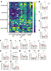
Results: Retinal arteriole and venule vasodilation was significantly increased in active + LIRD mice compared with inactive + LIRD mice. Superficial and intermediate/deep vascular plexi from inactive + LIRD mice had significantly decreased vessel density and total vessel length, with increased numbers of end points and lacunarity compared with active groups. Inactive + LIRD endothelial cells had significantly increased expression of VCAM1 and Nfkb1 compared with controls. Serum analyses revealed active + LIRD mice have a distinct immune response profile, with increased expression of keratinocyte-derived chemokine (KC), and VEGF-A compared to all other groups.

Conclusions: Treadmill exercise maintained retinal vascular morphology and function, altered endothelial gene expression, and is associated with a specific circulating immune response profile in the LIRD mouse model. These data support exercise as an effective intervention for maintaining retinal vascular integrity in a model of RD.


Conflict of interest statement

Disclosure: H.B. Haupt, None; J.M. Nickerson, None; J.H. Boatright, None; M.T. Pardue, None; K.L. Bales, None

Figures

Figure 1.
Figure 1.
Experimental methods and timeline. Six-week-old female BALB/c mice were assigned to inactive + dim, active + dim, inactive + LIRD, and active + LIRD groups (n = 20 per group). (A) Active mice were treadmill exercised (1 hour/day, 10 meters/minutes) for 3 weeks, and then, the LIRD was induced (5000 lux/4 hours). Inactive mice were placed on a static treadmill for the same schedule. (B) Retinal neurovascular coupling was measured by functional hyperemia. Eyes were illuminated using a 12 Hz flickering green light. (C) Scanning laser ophthalmoscope image identifying whether a vessel is a venule (Vx) or arteriole (Ax). (D) Plot of vessel diameter across time used to measure the percent change of vessel caliber during stimulation (highlighted region). (E) OCT-A vascular plexus segmentation with raw and processed images using Angiotool delineating superficial and intermediate/deep vascular plexi. Five days following LIRD, retinal extracts were pooled to perform (F) magnetic-activated cell sorting (MACS) to isolate retinal endothelial cells. The ddPCR was performed to quantify vascular gene expression. (G) Serum from all experimental groups was analyzed via multiplex assay. Figure was made with Biorender, available at https://BioRender.com/a73w831.
Figure 2.
Figure 2.
OCT-A reveals active   +   LIRD mice have maintained superficial vascular complex morphology. Optical coherence tomography angiography (OCT-A) was performed to evaluate superficial vascular complex morphology (SVC) in vivo (A–D). Inactive + LIRD mice displayed significant decreases in vessel density (E) and total vessel length (F) compared to active + dim animals, with no significant differences between inactive + dim and active + LIRD mice. However, inactive + LIRD mice had a significant increase in the total number of end points (G) compared with both active groups. Inactive + LIRD mice also had a significant increase in lacunarity (gaps, or regions without vasculature; H). Two-way ANOVA with Tukey's multiple comparison analyses were performed, graphs represent mean ± SEM, with each data point representing a single animal, n = 15, *P < 0.05, **P < 0.01,***P < 0.001.
Figure 3.
Figure 3.
Exercise promotes preservation of intermediate/deep vascular complex in mice undergoing light induced retinal degeneration. Optical coherence tomography angiography (OCT-A) was performed to evaluate intermediate/deep (I/D; A–D) vascular complex morphology in vivo. Inactive + LIRD mice displayed significant decreases in vessel density (E) and total vessel length (F) compared with the active groups and all groups, respectively. Active groups showed a significant decrease in the total number of end points compared to inactive groups (G). Inactive + LIRD mice also had a significant increase in lacunarity (gaps, or regions without vasculature; H). Two-way ANOVA with Tukey's multiple comparison analyses were performed, graphs represent mean ± SEM, with each data point representing a single animal, n = 15, *P < 0.05, **P < 0.01, ***P < 0.001, ****P < 0.0001.
Figure 4.
Figure 4.
Exercise protects against degradation of neurovascular coupling. Percent change of first order retinal arteriole and venule dilation was measured utilizing flicker induced functional hyperemia. (A–D) Representative plots of percent change in arteriole caliber over time, with blue highlighting the quantified region. Active + dim and active + LIRD mice had a significant increase in arteriole (E) and venule (F) dilation compared with inactive groups, with inactive + LIRD arteriole dilation being significantly decreased compared with all the groups. Two-way ANOVA with Tukey's multiple comparison analyses were performed, graphs represent mean ± SEM, with each data point representing a single animal, n = 10 to 13, *P < 0.05, **P < 0.01.
Figure 5.
Figure 5.
Inactive   +   LIRD endothelial cells have significantly increased expression of VCAM1 and Nfkb1 compared with controls. Isolated retinal endothelial cells from all experimental groups were probed for genes associated with angiogenesis, neuroprotection, and inflammation. Inactive + LIRD endothelial cells had significantly increased expression of vascular cell adhesion molecule 1 (VCAM1, C) expression compared with active + dim groups, whereas no significance was found between inactive + dim and active + LIRD groups. Inactive + LIRD endothelial cells also had significantly increased expression of nuclear factor kappa B subunit 1 (Nfkb1, G) compared with all groups, additionally active + dim animals were significantly decreased compared with active + LIRD groups. No statistical differences were observed in vascular endothelial growth factor receptor 1 (VEGFR1; A), VEGFR2 (B), endothelin-1 (EDN1; D) nitric oxide synthase 3 (NOS3, E), brain derived neurotrophic factor (BDNF, F), and nuclear factor kappa B subunit 2 (Nfkb2, H) expression. Two-way ANOVA with the two-stage Benjamin, Krieger, and Yekutieli procedure was performed, graphs represent mean ± SEM with each data point representing 5 animals (10 retinas), n = 4 per group, *P < 0.05.
Figure 6.
Figure 6.
Exercise and LIRD influence circulating cytokine and chemokine profiles. Multiplex analyses quantifying cytokine and chemokine expression related to circulating immune response. Active + LIRD animals have a specific circulating immune response profile compared to all other experimental groups, potentially supporting retinal neurons and promoting retinal vascular survival and function during retinal degeneration. Two-way ANOVA with Tukey's multiple comparison analyses were performed, graphs represent mean ± SEM values with each data point representing a single animal, n = 9 to 11 per group, *P < 0.05, **P < 0.01, ***P < 0.001.

Update of

References

    1. Davson H. The Bowman Lecture, 1979. The little brain. Trans Ophthalmol Soc U K (1962). 1979; 99(1): 21–37. - PubMed
    1. Ivanova E, Alam NM, Prusky GT, Sagdullaev BT. Blood-retina barrier failure and vision loss in neuron-specific degeneration. JCI Insight. 2019; 5(8): e126747. - PMC - PubMed
    1. Otani A, Dorrell MI, Kinder K, et al.. Rescue of retinal degeneration by intravitreally injected adult bone marrow-derived lineage-negative hematopoietic stem cells. J Clin Invest. 2004; 114(6): 765–774. - PMC - PubMed
    1. Böhm EW, Pfeiffer N, Wagner FM, Gericke A. Methods to measure blood flow and vascular reactivity in the retina. Front Med (Lausanne). 2023; 9: 1069449. - PMC - PubMed
    1. Taylor TRP, Menten MJ, Rueckert D, Sivaprasad S, Lotery AJ. The role of the retinal vasculature in age-related macular degeneration: a spotlight on OCTA. Eye (Lond). 2024; 38(3): 442–449. - PMC - PubMed

LinkOut - more resources