Review
. 2025 Aug 26;18(1):45.
doi: 10.1007/s40820-025-01890-2.

Wearable Ultrasound Devices for Therapeutic Applications

Affiliations
Review

Wearable Ultrasound Devices for Therapeutic Applications

Sicheng Chen et al. Nanomicro Lett. .

Abstract

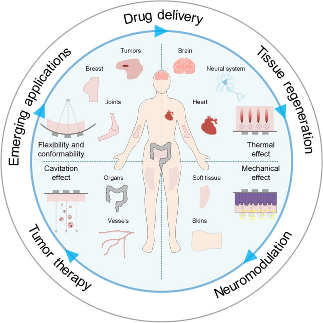
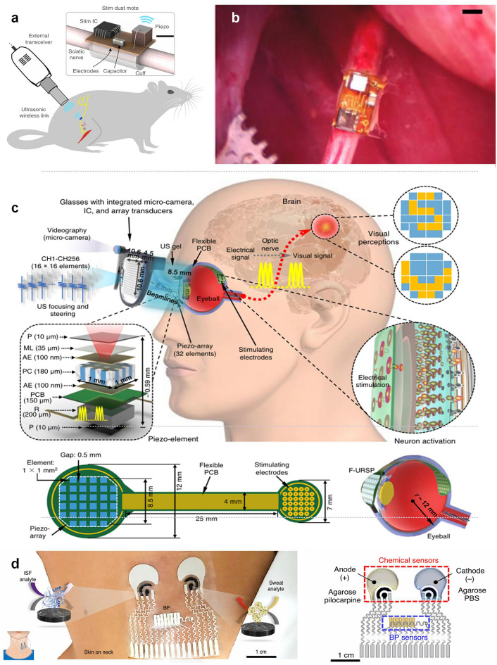
Wearable ultrasound devices represent a transformative advancement in therapeutic applications, offering noninvasive, continuous, and targeted treatment for deep tissues. These systems leverage flexible materials (e.g., piezoelectric composites, biodegradable polymers) and conformable designs to enable stable integration with dynamic anatomical surfaces. Key innovations include ultrasound-enhanced drug delivery through cavitation-mediated transdermal penetration, accelerated tissue regeneration via mechanical and electrical stimulation, and precise neuromodulation using focused acoustic waves. Recent developments demonstrate wireless operation, real-time monitoring, and closed-loop therapy, facilitated by energy-efficient transducers and AI-driven adaptive control. Despite progress, challenges persist in material durability, clinical validation, and scalable manufacturing. Future directions highlight the integration of nanomaterials, 3D-printed architectures, and multimodal sensing for personalized medicine. This technology holds significant potential to redefine chronic disease management, postoperative recovery, and neurorehabilitation, bridging the gap between clinical and home-based care.

Keywords: Closed-loop therapy; Drug delivery; Neurorehabilitation; Tissue regeneration; Wearable ultrasound devices.


Conflict of interest statement

Declarations. Conflicts of interest: The authors declare no interest conflict. They have no known competing financial interests or personal relationships that could have appeared to influence the work reported in this paper.

Figures

Fig. 1
Fig. 1
Summary of ultrasound technology applications across diverse therapeutic purposes, including drug delivery, tissue regeneration, neuromodulation, and tumor therapy. Wearable biodevices demonstrate emerging applications tailored to specific organs
Fig. 2
Fig. 2
a The range of ultrasonic wave frequencies and the relationship between attenuation coefficient and frequency [54]. Copyright 2018, IEEE. b Overview of relevant ultrasound wave characteristics. The ultrasound wave characteristics strongly define microbubble cavitation behavior and biological outcome and are therefore important to report. Reproduced with permission [56]. Copyright 2021, Elsevier
Fig. 3
Fig. 3
a Exploded view of the 2D-array of cUSP showing each constituent layer; scale bar: 5 mm (the inset shows the fluid cavity formed between the PZT-D element and skin; scale bar: 2 m. Reproduced with permission [88]. Copyright 2023, Wiley–VCH GmbH. b Broadband noise amplitude as a function of applied voltage to the PZT-D. Reproduced with permission [88]. Copyright 2023, Wiley–VCH GmbH. c Simulated acoustic emission profile of a piezoelectric material with a size of 0.2 × 0.2 mm2 (inset) with excellent beam directivity, focus, and penetration depth (> 40 m. Reproduced with permission [89]. Copyright 2024, Wiley–VCH GmbH. d Relative coverage percentages of PCNA- and TUNEL- positive areas. Data are expressed as mean ± SD (n = 5); The number of TUNEL- positive areas between group 1 and group 3; The number of TUNEL- positive areas between group 2 and group 3; The differences among groups were calculated using one- way ANOVA. *p < 0.05, **p < 0.01. Reproduced with permission [89]. Copyright 2024, Wiley–VCH GmbH. e Sound waves are distributed vertically along the direction of the array when the bending degree ranges from 0 to 45 degree. Reproduced with permission [90]. Copyright 2021, Wiley–VCH GmbH
Fig. 4
Fig. 4
a PMMA, PI and Cu metal layers are spin-coated on the Si substrate. Lithography, etching and transfer printing are used to fabricate the flexible circuit. Piezoelectric ultrasonic units (PZT—4) are integrated into the flexible circuit, which is then packaged with a thin (≈200 μ hydrogel patch. Reproduced with permission [90]. Copyright 2021, Wiley–VCH GmbH. b Optical image (top view) of four elements, showing the morphology of the backing layers and top electrodes, and the optical image (bottom view) of four elements, showing the morphology of the piezoelectric material and bottom electrodes. Reproduced with permission [34]. Copyright 2018, AAAS. c Bent around a developable surface. Reproduced with permission [31]. Copyright 2018, Springer Nature. d Compression test of the hybrid NG: short circuit current and open circuit voltage. Reproduced with permission [118]. Copyright 2020, WILEY–VCH Verlag GmbH & Co. KGaA
Fig. 5
Fig. 5
a Comparison of calibration curves for a biodegradable sensor using stretched, bulk piezo PLLA film (bl and a 4,000 rpm electrospun PLLA nanofiber film (red). Reproduced with permission [120]. Copyright 2019, PNAS. b Output from a charge amplifying circuit connected to a 4,000 rpm electrospun, biodegradable PLLA sensor that is subjected to 10,000 cycles of a 10-N force. Reproduced with permission [120]. Copyright 2019, PNAS. c Comparison of the simulated abdominal pressure signals, wirelessly recorded from an implanted biodegradable PLLA nanofiber sensor using a 300 rpm negative control (bland a 4,000 rpm film). Reproduced with permission [120]. Copyright 2019, PNAS. d Schematic illustration of glycine-PCL nanofibers. Reproduced with permission [121]. Copyright 2023, AAAS. e Kaplan–Meier survival of animals receiving different treatments (n = 8, log-rank test). Reproduced with permission [121]. Copyright 2023, AAAS. f Conductivity as a function of nozzle diameter for 3D-printed conducting polymers in dry and hydrogel states. Reproduced with permission [122]. Copyright 2020, Springer Nature. g 3D-printed conducting polymers can be converted into a pure PEDOT:PSS both in dry and hydrogel states by dry-annealing and subsequent swelling in wet environment, respectively. Reproduced with permission [122]. Copyright 2020, Springer Nature
Fig. 6
Fig. 6
a Ultrasonic transmission using a few ultrasonic transducers.Ultrasonic transmission by a tiled array of ultrasonic transducers. Reproduced with permission [97]. Copyright 2021, AAAS. b Ultrasonic transmission by an array of ultrasonic transducers placed inside the top part of a flexible base. Reproduced with permission [97]. Copyright 2021, AAAS. c USD-TENG system is implanted under the skin and receives wireless energy from ultrasound to power the implanted IMDs. Reproduced with permission [131]. Copyright 2022, Elsevier Inc
Fig. 7
Fig. 7
a Diagram of the StimDust system. Here it is used to stimulate the sciatic nerve of a rat. Scale bar. Reproduced with permission [133]. Copyright 2020, Springer Nature. b Stimulating mote fully implanted and affixed to a rat sciatic nerve. Reproduced with permission [133]. Copyright 2020, Springer Nature. c Schematic diagram showing the flexible ultrasonic device, with key components and aspect ratios labeled. Reproduced with permission [36]. Copyright 2022, Springer Nature. d Illustrations of the placement of the sensor and the enzymatic chemical sensors for ISF and sweat. Reproduced with permission [134]. Copyright 2021, Springer Nature
Fig. 8
Fig. 8
a Schematic of the integrated system level wf-UMP electronics, consisting of a flexible US transducer array for effective US emission, a bioadhesive hydrogel elastomer for robust adhesion and acoustic coupling layer, and a mKNN PNPs-loaded MN patch for drug delivery. Reproduced with permission [140]. Copyright 2025, Springer Nature. b FCM results of CD3 + CD8 + T cells and CD3 + CD4 + T cells infiltrated in tumors. Reproduced with permission [140]. Copyright 2025, Springer Nature. c Thermal images of wf-UMP on an agarose hydrogel operated continuously for 30 min. Top, 0 min. Bottom: 30 min. Scale bars, 1 cm. Reproduced with permission [141]. Copyright 2023, Springer Nature. d Comparison of drug delivery perfor mance of wf-UMP on agarose hydrogels with/without US stim ulation. Scale bars, 2 mm. Reproduced with permission [141]. Copyright 2023, Springer Nature. e Confocal microscopy images of porcine skin treated with Rho B labeled wf UMP with/without US stimulation at a depth of 328 µm. Scale bars, 250 µm. Reproduced with permission [141]. Copyright 2023, Springer Nature. f Quantifica tion of in vivo RhB dye release using the different methods. Reproduced with permission [141]. Copyright 2023, Springer Nature. g Presence of RhB dye in mouse muscle, liver, and kidney 24 h after the indicated treatments. Scale bar, 200 µm. Reproduced with permission [141]. Copyright 2023, Springer Nature. h After acoustic metamaterial treatments, representative images of mouse skin indi cated acoustic-mediated RhB dye delivery for 30, 60, or 180 s (fluorescent images). Reproduced with permission [141]. Copyright 2023, Springer Nature. i IVISR images of mice post dye delivery (10, 60, or 240 mvia acoustic metamaterials methods. Reproduced with permission [141]. Copyright 2023, Springer Nature. j Multiphoton confocal microscopy image showing RhB (rhodamin penetration into a vertical Sect. (7 μm thi of a porcine skin sample following passive diffusion; scale bar: 200 μm. Reproduced with permission [88]. Copyright 2023, Wiley–VCH GmbH. k Schematic illustration of the cUSP on skin, showing the cavitation mechanism within the cavity between the device and the skin, and resulting drug penetration through stratum corneum. Reproduced with permission [88]. Copyright 2023, Wiley–VCH GmbH. l Amount of NIA permeated with application of ultrasound versus control (passive permeafor the cUSP array. Reproduced with permission [88]. Copyright 2023, Wiley–VCH GmbH
Fig. 9
Fig. 9
a Schematic of the BA-TENG in sealing and ultrasound driven electrically healing rat skin injuries. Reproduced with permission [144]. Copyright 2023, Wiley–VCH GmbH. b Voltage and current output of the BA-TENG measured in water at 5∼mm from the ultrasound probe under 1∼W cm−2. Reproduced with permission [144]. Copyright 2023, Wiley–VCH GmbH. c Comparison of 3-day wound healing images in the untreated (contsuture, bioadhesive PAV only, and BA-TENG treated groups. Scalebar:1∼cm. Reproduced with permission [144]. Copyright 2023, Wiley–VCH GmbH. dSchematic diagram of wound treatment with the flexible ultrasonic patch. Reproduced with permission [90]. Copyright 2021, Wiley–VCH GmbH. e Healing states of the wound surface during treatment for the eight groups. Reproduced with permission [90]. Copyright 2021, Wiley–VCH GmbH. f Conceptual illustration of multifunctional UEP in high-quality wound healing and monitoring. Reproduced with permission [145]. Copyright 2025, Wiley–VCH GmbH. g Optical images of wound closure collected on days 0, 4, 8, 12, and 16 for the control, US, LUE, and HUE groups. Reproduced with permission [145]. Copyright 2025, Wiley–VCH GmbH. h 3D color mapping of skin–wound resistance trends (named: recovery cur. The prediction of wound monitoring and healing time under the recovery curve. Reproduced with permission [145]. Copyright 2025, Wiley–VCH GmbH
Fig. 10
Fig. 10
a Schematic diagram of the piezoelectric nanofiber scaffolds combining US promoted NSCs differentiation. Reproduced with permission [150]. Copyright 2022, ACS Nano. b Ultrasound powered wireless ES based on a biodegradable high-performance 3D piezoelectric scaffold promotes neural regeneration in spinal cord injuries. Reproduced with permission [150]. Copyright 2022, ACS Nano. c Immunofluorescent stain ing of the early neural markers Nestin and β-Tubulin III (Tuj1), the later neural marker MAP2, and neurogliocyte specific maker glial fibrillary acidic protein (GFAP) after 7 days culture. Reproduced with permission [150]. Copyright 2022, ACS Nano. d Statistical analysis of the f luorescence intensity of Nestin. Reproduced with permission [150]. Copyright 2022, ACS Nano. e Relative mRNA expression of four proteins. Reproduced with permission [150]. Copyright 2022, ACS Nano. f Off-center beam steering, dynamic beam steering along the wave- propagation direction by adjusting the ultrasound frequency without altering the metasurface, and dual focusing. Reproduced with permission [151]. Copyright 2024, PNAS. g Assessment of mouse immobility, quantified as the percentage of time spent immobile before, during, and after ultrasound stimulation. Reproduced with permission [151]. Copyright 2024, PNAS. h Observed enhancement in motor activity demonstrates the therapeutic potential of bilateral AhSonogenetic stimulation in restoring function in neurodegenerative conditions. Reproduced with permission [151]. Copyright 2024, PNAS

References

    1. H. Wu, W. Gao, Z. Yin, Materials, devices and systems of soft bioelectronics for precision therapy. Adv. Healthc. Mater. 6(10), 1700017 (2017). 10.1002/adhm.201700017 - PubMed
    1. M. Lin, H. Hu, S. Zhou, S. Xu, Soft wearable devices for deep-tissue sensing. Nat. Rev. Mater. 7(11), 850–869 (2022). 10.1038/s41578-022-00427-y
    1. H.C. Ates, P.Q. Nguyen, L. Gonzalez-Macia, E. Morales-Narváez, F. Güder et al., End-to-end design of wearable sensors. Nat. Rev. Mater. 7(11), 887–907 (2022). 10.1038/s41578-022-00460-x - PMC - PubMed
    1. J. Chen, Y. Zhu, X. Chang, D. Pan, G. Song et al., Recent progress in essential functions of soft electronic skin. Adv. Funct. Mater. 31(42), 2104686 (2021). 10.1002/adfm.202104686
    1. J. Heikenfeld, A. Jajack, J. Rogers, P. Gutruf, L. Tian et al., Wearable sensors: modalities, challenges, and prospects. Lab Chip 18(2), 217–248 (2018). 10.1039/C7LC00914C - PMC - PubMed

LinkOut - more resources