. 2025 Sep 2;122(35):e2426145122.
doi: 10.1073/pnas.2426145122. Epub 2025 Aug 26.

Ovarian germline stem cell dedifferentiation is cytoneme dependent

Affiliations

Ovarian germline stem cell dedifferentiation is cytoneme dependent

Catherine Sutcliffe et al. Proc Natl Acad Sci U S A. .

Abstract

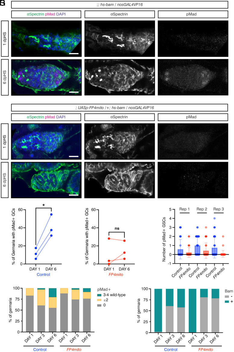
Progenitor cell dedifferentiation is important for stem cell maintenance during tissue repair and age-related stem cell decline. Here, we use the Drosophila ovary as a model to study the role of cytonemes in bone morphogenic protein (BMP) signaling-directed germline stem cell (GSC) maintenance and dedifferentiation of germ cells to GSCs. We provide evidence that differentiating germ cell cysts extend longer cytonemes that are more polarized toward the niche during dedifferentiation to reactivate BMP signaling. The presence of additional somatic cells in the niche is associated with a failure of germ cell dedifferentiation, consistent with the formation of a physical barrier to cytoneme-niche contact and outcompetition of germ cells for BMP. Using BMP beads in vitro, we show that these are sufficient to induce cytoneme-dependent contacts in Drosophila tissue culture cells. We demonstrate that the Enabled (Ena) actin polymerase is localized to the tips of germ cell cytonemes and is necessary for robust cytoneme formation, as its mislocalization reduces the frequency, length, and directionality of cytonemes. During homeostasis, specifically perturbing cytoneme function through Ena mislocalization impairs GSC fitness by reducing GSC BMP signaling and niche occupancy. Disrupting cytonemes by targeting Ena during dedifferentiation reduces germ cell BMP responsiveness and the ability of differentiating cysts to dedifferentiate. Overall, our results provide evidence that cytonemes play a fundamental role in establishing polarized signaling and niche occupancy during stem cell maintenance and dedifferentiation.

Keywords: BMP signaling; cytoneme; dedifferentiation; enabled; germline stem cell.


Conflict of interest statement

Competing interests statement:The authors declare no competing interest.

Figures

Fig. 1.
Fig. 1.
GCs dedifferentiate over time to reoccupy the niche. (A) Schematic of the germarium with the different cell types, as labeled, anterior is to the Left. The spectrosome and fusomes are shown in white. The boxed germarium shows post-HS induced bam expression. (BG) Germaria carrying bam promoter-GFP and hs-bam transgenes immunostained with anti-pMad (magenta), anti-GFP (green), anti-αSpectrin (gray, Right panel), and DAPI (blue). Representative germaria are shown for day 0 (B) and 1 dpHS (C), 3 dpHS (D and E), and 6 dpHS (F and G). (*) Cap cells. (Scale bar, 10 μm.) (H) Quantitation of the number of germaria where the GCs contacting the niche are pMad-positive or -negative differentiating cysts or pMad-positive GSCs on 0, 1, 3, and 6 dpHS. The number of germaria counted per time point was 104, 100, 101, and 103 for 0, 1, 3, and 6 dpHS, respectively. (I and J) pMad-positive cysts at 3 dpHS immunostained with anti-pMad (magenta, gray Right panel), anti-αSpectrin (green), and DAPI (blue). The image in (I) is from the germarium shown in (E). Connected CBs are outlined and numbered 1 to 4 in the 4-cell cyst. (*) Cap cells, the arrowhead shows a fusome undergoing scission. (Scale bar, 5 μm.)
Fig. 2.
Fig. 2.
Dedifferentiating GCs extend cytonemes during niche recolonization. (AC) Germaria carrying UASp-LifeAct-GFP driven by nosGAL4VP16 and an hs-bam transgene immunostained with anti-GFP (green and Inset), anti-αSpectrin (magenta), anti-pMad (Inset), and DAPI (blue). The right panels show higher magnification views of the boxed areas. Non-HS control (A), 3 dpHS germaria (B and C) with (B) a pMad negative 8 cc extending cytonemes to the niche and (C) a pMad-positive GSC with cytonemes contacting the niche. (*) Niche cells including extra somatic cells, cyan arrowheads mark cytonemes, magenta arrowheads indicate pMad-positive cells, white dotted lines outline pMad-positive GSCs. (Scale bars, 5 μm.) (D) Graph shows quantitation of the number of niche cells in hs-bam germaria (3 dpHS) that contain pMad-positive dedifferentiating GCs or only pMad-negative GCs, compared to control germaria (HS, with no hs-bam transgene). n = 30, data shown as mean ± SD, ANOVA P =< 0.001 (control vs. pMad− GCs), P =< 0.001 (pMad+ GCs vs. pMad− GCs). (E and F) Images of live germaria carrying UASp-LifeAct-tagRFP driven by nosGAL4VP16 (magenta and Inset), a bamP-GFP reporter to mark differentiating cells (green and Inset), and an hs-bam transgene. Non-HS control (E) and 3 dpHS germaria (F) show the cytonemes on a GSC and CBs (E, arrowheads) or a CB occupying the niche (F, arrowheads). Higher magnification views of LifeAct-tagRFP labeled CB cytonemes are shown in the Right panel. (*) Niche cells. (Scale bar, 5 μm and Inset 2μm.) (GI) Quantification of the number of cytonemes per CB (G), cytoneme length (H), and orientation relative to the niche (0°), each interval is 10% (I). n = 50 (hs-bam) and 46 (control), Welch’s unpaired two-tailed t test, P < 0.0001, ns not significant. (J) Schematic of the germarium niche showing no HS GSCs and a 3 dpHS cyst which extends cytonemes toward the niche and is BMP responsive. (K and L) S2R+ cells transfected with Tkv-mCherry and FLAG-Mad expression plasmids and incubated with BSA (K) and BMP2 (L) coated beads. S2R+ cells were immunostained with anti-tdTomato (magenta and Inset) and anti-pMad (green and Inset). Bead locations are marked with yellow dashed lines. Brightfield images and higher magnification views of the Tkv-mCherry channel are shown. (Scale bar, 10 μm and Inset 2μm.) (M) Quantification of the contacts between BSA and BMP2-coated beads and S2R+ cells (examples are shown in K and L). n = 45, Fisher’s exact test, P = 0.0071. (N) Quantification of the distance between the bead and the center of the cell nucleus. Mann–Whitney P = 0.0781.
Fig. 3.
Fig. 3.
Ena is localized to cytoneme tips. (A) Wildtype germarium immunostained with anti-Ena (magenta), Phalloidin (green), and DAPI (blue). GSCs and CpCs are marked by brackets. (Scale bar, 10 μm.) (B) Immunostaining as in (A) and Vasa (gray Inset). A GSC is outlined by a dotted line based on Vasa staining, white arrowheads mark cytonemes (i and ii), that are shown as higher magnification images. (*) Cap Cells. [Scale bar, 5 μm and (i-ii) 1 μm.] (C) Immunostaining as in (B). A GSC and CB are outlined by dotted lines. A CB cytoneme is indicated by a white arrowhead and shown magnified in (i). Lamellipodial projections are indicated by brackets. (*) Cap Cells. [Scale bar, 5 μm and (i) 1 μm.] (D and E) Images of a GSC (D) and CB (E) from live germaria expressing LifeAct-tagRFP (magenta and Inset) and Ena-GFP (green and Inset) driven by nosGAL4VP16. Higher magnification views of cytonemes are shown. (*) Cap Cells. (Scale bar, 5 μm and Inset 1 μm.) (F) Image from a live germarium expressing GAP43-Venus driven by nosGAL4VP16. A GSC and CB are shown, with higher magnification views of the cytonemes. (*) Cap Cells. (Scale bar, 5 μm and Inset 1 μm.) (G) A GSC imaged in a fixed germarium expressing GAP43-Venus driven by nosGAL4VP16 and immunostained with anti-GFP (green and Inset), anti-Ena (magenta and Inset), and DAPI (blue). The higher magnification view shows a cytoneme. (*) Cap Cells. (Scale bar, 5 μm and Inset 1 μm). (H) Wildtype germarium immunostained with anti-Ena (magenta), anti-Ecad (green), and DAPI (blue). The GSC is outlined by a dotted line. (*) Cap Cells. (Scale bar, 5 μm and Inset 1 μm.) (I) Intensity plot for Ena and Ecad staining shown in (H) across points 1 to 4 of the CpC marked with a yellow asterisk. (J) Control and FP4mito expressing germaria immunostained with anti-Ecad (green and gray Inset) and DAPI (blue). (*) Cap Cells. (Scale bar, 5 μm.) (K) Intensity plot for Ecad staining, as shown in (J), n = 29 and 23, Welch’s unpaired two-tailed t test, P = 0.5942 (ns).
Fig. 4.
Fig. 4.
Ena regulates GC cytoneme formation. (A and B) Live images of control and FP4mito expressing germaria, with LifeAct-tagRFP (magenta and Inset) and Ena-GFP (green and Inset) driven by nosGAL4VP16. Images show a GSC (A) or CB (B), with higher magnification views of a cytoneme. (*) Cap Cells. (Scale bar, 5 μm.) (C and D) Graphs showing the number of cytonemes per GSC or CB and cytoneme lengths from live control and FP4mito germaria, as shown in (A and B) for representative examples. n = 11-13 cytonemes, Welch’s unpaired t test, ****P =< 0.0001 and **P =< 0.01. (E and F) Data from (C and D) showing the number of cytonemes per GC projecting either anteriorly or laterally from live control and FP4mito germaria, as shown in (A and B) for representative examples. Welch’s unpaired t test, *P =< 0.05.
Fig. 5.
Fig. 5.
Ena mislocalization impairs BMP signal reception and stem cell fitness. (A and B) Representative images of control or FP4mito germaria aged for 7 d, immunostained with anti-αSpectrin (gray), anti-pMad (magenta and Inset), anti-Bam (green and Inset), and DAPI (blue). Magenta arrowheads indicate pMad-positive GCs. (Scale bar, 10 μm.) (C) Graph shows the number of pMad-positive GCs counted in 7-d-old germaria including the representative germaria shown in (A and B). n = 50. (D) Graph shows the pMad intensity in GSCs in control and FP4mito backgrounds. n = 34 per genotype, data shown as mean ± SD, Welch’s two-tailed unpaired t test, P = 0.0003 for comparisons between genotypes. (E) Graph shows the proportion of germaria with and without Bam-positive cysts counted in 7-d-old samples. n = 50. (F and G) As in (A and B), except the germaria were aged for 2.5 wk. (H) Graph shows the number of pMad-positive GCs counted in 2.5-wk-old germaria, including the representative germaria from (F and G). n = 50. (I) Graph shows the proportion of germaria with and without Bam-positive cysts counted in 2.5-wk-old samples. n = 50.
Fig. 6.
Fig. 6.
Ena mislocalization impairs GSC dedifferentiation. (A and B) Control germaria carrying an hs-bam transgene and nosGAL4VP16 immunostained with anti-pMad (magenta and Inset), anti-αSpectrin (green and Inset), and DAPI (blue). Representative germaria are shown for day 1 (A) and 6 (B) post-HS. (Scale bar, 10 μm.) (C and D) As in (A and B), except germaria also carry a UASp-FP4mito transgene. (Scale bar, 10 μm.) (E and F) Quantification of germaria containing pMad-positive early GCs on days 1 and 6 post-HS for (E) control and (F) FP4mito. Data are pairwise comparisons, two-tailed paired t test comparing day 1 to day 6, P = 0.0150 for control (*) and P = 0.448 for FP4mito (ns). Each repeat contains at least 30 germaria. (G) Quantification of the number of pMad-positive GSCs per germaria for each repeat. Data are shown as mean ± SD. (H and I) Graph shows the number of pMad-positive GCs (H) or Bam-positive GCs (I) counted in 1, 3, and 6 dpHS (representative examples are in SI Appendix, Fig. S5 AJ).
Fig. 7.
Fig. 7.
Cytonemes promote stem cell fitness and enable cyst dedifferentiation. Model of the role of cytonemes in the ovarian germline at homeostasis, in promoting Dpp signaling and stem cell fitness, and in dedifferentiation, by enabling GCs to access niche-derived Dpp.

References

    1. Rojas-Rios P., Gonzalez-Reyes A., Concise review: The plasticity of stem cell niches: A general property behind tissue homeostasis and repair. Stem Cells 32, 852–859 (2014). - PubMed
    1. Ramirez-Weber F. A., Kornberg T. B., Cytonemes: Cellular processes that project to the principal signaling center in Drosophila imaginal discs. Cell 97, 599–607 (1999). - PubMed
    1. Daly C. A., Hall E. T., Ogden S. K., Regulatory mechanisms of cytoneme-based morphogen transport. Cell Mol. Life Sci. 79, 119 (2022). - PMC - PubMed
    1. Ramesh P., Dey N. S., Kanwal A., Mandal S., Mandal L., Relish plays a dynamic role in the niche to modulate Drosophila blood progenitor homeostasis in development and infection. Elife 10, e67158 (2021). - PMC - PubMed
    1. Patel A., et al. , Cytonemes coordinate asymmetric signaling and organization in the Drosophila muscle progenitor niche. Nat. Commun. 13, 1185 (2022). - PMC - PubMed

Substances

LinkOut - more resources