. 2025 Aug 22;10(16):e190534.
doi: 10.1172/jci.insight.190534.

Single-cell spatial transcriptomics unravels the cellular landscape of abdominal aortic aneurysm

Affiliations

Single-cell spatial transcriptomics unravels the cellular landscape of abdominal aortic aneurysm

Guizhen Zhao et al. JCI Insight. .

Abstract

Abdominal aortic aneurysm (AAA) is a life-threatening vascular disease with no effective pharmacological interventions. While single-cell transcriptomics has advanced our understanding of AAA, it lacks spatial context. Here, we employed Seq-Scope, an ultra-high-resolution spatial transcriptomic technology, to decipher the spatial landscape of angiotensin II-induced AAA in Apoe-/- mice. Our analysis revealed the heterogeneity of macrophages, fibroblasts, and smooth muscle cells (SMCs), with specific responses in different layers of the AAA tissue. SMCs in the inner layers showed associations with Mgp-expressing fibroblasts and GPNMB-expressing macrophages, whereas the outer layers had different dominant cell types. Notably, GPNMB-expressing macrophages were concentrated near SMCs in regions of severe elastic lamina damage. Immunofluorescent staining confirmed their colocalization, and scRNA-seq reanalysis independently validated the presence of GPNMB-high macrophages in AAA tissues, highlighting their involvement in inflammation and tissue remodeling. Moreover, we discovered that macrophage-derived soluble GPNMB induces SMC phenotypic switching, reducing contractile markers while increasing cytokines and metalloproteinases. This effect was partly mediated by CD44 signaling. These findings suggest that GPNMB-high macrophages contribute to AAA development by driving SMC dysfunction. This study highlights the importance of high-resolution spatial transcriptomics in complementing single-cell transcriptomics, offering valuable insights into molecular and cellular responses in the AAA microenvironment.

Keywords: Cardiology; Cardiovascular disease; Vascular biology.


Figures
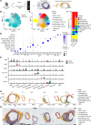
Figure 1
Figure 1. Single-cell spatial transcriptomic profiling of normal and aneurysmal abdominal aortas by Seq-Scope.
Sixteen-week-old male Apoe–/– mice were infused with AngII (1,000 ng/kg/min) or saline via minipumps for 4 weeks. n = 5/group. (A) Schematic diagram depicting the mouse abdominal aortic aneurysm (AAA) and the spatial-transcriptomic analysis. (B) Uniform manifold approximation and projection (UMAP) of spatial transcriptomics spots from normal control and aneurysm group. The transcriptomics spots were from 10 sections of 3 normal control and 10 sections of 3 AAA group. (C) UMAP of cell-type clusters based on the gene compositions in each spatial transcriptomics spot. SMC, smooth muscle cells; Csrp2, SMC_Csrp2, SMCs highly expressing cysteine and glycine-rich protein 2; Fibroblast_Col, fibroblasts highly expressing collagen; Fibroblast_Mgp, fibroblasts highly expressing matrix Gla protein; Firboblast_Tmem119, fibroblast highly expressing transmembrane protein 119; Macrophage_Ctsb, macrophages highly expressing cathepsin B; Macrophage_Gpnmb, macrophages highly expressing glycoprotein Nmb; RBC, red blood cell. (D) Dot plot of marker genes for each cell population. (E) StackedVlnPlot showing the expression of marker genes across Control and Aneurysm groups. (F) Cell population percentages in each group. (G and H) Representative spatial plots of cell-type clusters (G) were overlaid with H&E imaging (H).
Figure 2
Figure 2. Spatial mapping of macrophages, GPNMB-high macrophages, and SMCs in normal aorta and AAA.
(A) Spatial plot showing the spatial prediction score of SMC, SMC_Csrp2, Macrophage_Gpnmb, and Fibroblast_Col in the aortic tissues. (B and C) Spatial plot visualizing locations of SMC, SMC_Csrp2, Macrophage_Gpnmb, and Fibroblast_Col in the tissue sections. The boxed areas (blue boxes) are magnified on the right.
Figure 3
Figure 3. Immunofluorescent staining confirms the interaction of SMC and GPNMB-high macrophages in the aneurysmal aorta.
(A) Immunofluorescent staining of GPNMB (red), Mac2 (cyan), and α-actin (green) to respectively show SMCs, macrophages, and GPNMB+ cells in the aortic wall of control and AAA. Nuclei stained with DPAI are blue. Scale bars: 100 μm (whole sections) and 50 μm (higher magnification areas). (B) Quantification of the GPNMB, Mac, and α-actin fluorescence intensity. (C) Correlation analysis between GPNMB and Mac2 or α-actin in AAA groups. Data are presented as mean ± SEM. *P < 0.05 by 2-tailed Student’s t test.
Figure 4
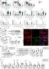
Figure 4. Macrophage-secreted GPNMB promotes SMC phenotypic switching.
(A) Violin plot of Myh11, Tagln, Csrp2, Cd68, Lyz2, Gpnmb, Cd44, Egfr, Kdr, Mmp2, and Mmp9 expression in SMC and macrophage subpopulations. The results were reanalyzed from scRNA-seq data (GSE193265 and GSE191226). The single cells were from healthy suprarenal abdominal aorta (Control) and abdominal aortic aneurysm (AAA) isolated from male ApoE–/– mice. (BD) BMDMs isolated from C57BL/6J mice were stimulated with or without 10 ng/mL TNF-α for 24 hours. (B and C) Gpnmb mRNA and protein levels were determined. (D) The GPNMB protein abundance in the culture media was determined by ELISA (n = 6). (E and F) Human aortic smooth muscle cells (HASMCs), at 80% confluence, were serum starved for 24 hours and then treated with 40 ng/mL recombinant GPNMB (aa 22–486, sGPNMB) protein in fresh Opti-MEM for 48 hours. (E) qPCR was performed to determine the mRNA levels of GPNMB, GPNMB receptors CD44 and KDR, and protease ADAM10 (n = 6). (F) Immunofluorescent staining of SM22α and CD44. Nuclei stained with DPAI are blue. Scale bars: 20 μm. (GI) HASMCs were transfected with nontargeting control siRNA (siControl) or siCD44. After 24 hours, the cells were serum starved for 24 hours and then treated with sGPNMB for 48 hours. qPCR was performed to determine the mRNA levels of CD44 (G, upper panel, n = 6), SMC contractile markers MYH11, ACTA2, and TAGLN and inflammatory cytokines CCL2 and IL6 as well as proteases MMP2, MMP9, and ADAM10 (H, n = 6). The protein abundance of CD44 (G, lower panel, n = 3) smooth muscle α-actin, calponin, and SM22α (I, n = 6) was determined by Western blot. Data are presented as mean ± SEM. Wilcoxon’s test was used for comparing means in A, 2-tailed Student’s t test for B, D, and E, and 1-way ANOVA followed by Tukey’s post hoc test for GI. *P < 0.05.

References

    1. Nordon IM, et al. Pathophysiology and epidemiology of abdominal aortic aneurysms. Nat Rev Cardiol. 2011;8(2):92–102. doi: 10.1038/nrcardio.2010.180. - DOI - PubMed
    1. Lu H, et al. Cyclodextrin prevents abdominal aortic aneurysm via activation of vascular smooth muscle cell transcription factor EB. Circulation. 2020;142(5):483–498. doi: 10.1161/CIRCULATIONAHA.119.044803. - DOI - PMC - PubMed
    1. Zhao G, et al. BAF60c prevents abdominal aortic aneurysm formation through epigenetic control of vascular smooth muscle cell homeostasis. J Clin Invest. 2022;132(21):e158309. doi: 10.1172/JCI158309. - DOI - PMC - PubMed
    1. Davis FM, et al. Single-cell transcriptomics reveals dynamic role of smooth muscle cells and enrichment of immune cell subsets in human abdominal aortic aneurysms. Ann Surg. 2022;276(3):511–521. doi: 10.1097/SLA.0000000000005551. - DOI - PMC - PubMed
    1. Yang H, et al. Single-cell RNA sequencing reveals heterogeneity of vascular cells in early stage murine abdominal aortic aneurysm-brief report. Arterioscler Thromb Vasc Biol. 2021;41(3):1158–1166. doi: 10.1161/ATVBAHA.120.315607. - DOI - PMC - PubMed

MeSH terms