. 2025 Sep;4(9):1094-1113.
doi: 10.1038/s44161-025-00691-5. Epub 2025 Aug 26.

Inhibition of proprotein convertase SKI-1 prevents blood vessel alteration after stroke

Affiliations

Inhibition of proprotein convertase SKI-1 prevents blood vessel alteration after stroke

Alireza P Shabanzadeh et al. Nat Cardiovasc Res. 2025 Sep.

Abstract

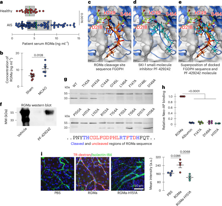
Neutralizing factors involved in blood vessel dysfunction offer a promising strategy for stroke recovery. Many extracellular proteins need enzymatic activation to function, and blocking this activation is an untapped approach to restoring vessel integrity. Here we demonstrate that inhibition of the extracellular protease SKI-1 with PF-429242 restores blood vessel integrity and promotes functional recovery in both large and small animal models for stroke. Single-cell mRNA sequencing identified molecular signatures suggesting that PF-429242 restores the expression of genes involved in vessel integrity in endothelial cells. Moreover, we identify a mechanism whereby RGMa cleavage by SKI-1 is required for RGMa to interact with Neogenin and alter vessel integrity. Either preventing RGMa cleavage or deleting Neogenin on endothelial cells reduced blood vessel dysfunction, increased tissue preservation and restored brain function after stroke. This work identifies a much-needed therapeutic strategy that restores blood vessel integrity and functionality, showing efficacy in large and small animals.


Conflict of interest statement

Competing interests: The authors declare no competing interests.

Figures

Fig. 1
Fig. 1. Gene expression signatures associated with PF-429242 rescue.
a, Schematic of the protocol used to study the role of SKI-1 in stroke: mice received MCAO followed by treatment with PF-429242 or PBS. Two days after MCAO, brains were collected and dissociated into single cells; endothelial cells were isolated using FACS; and mRNA sequencing of single cells was performed and analyzed (https://biorender.com/ytb0wjd). bg, Expression overlaid t-SNE plots of gene markers (intensity indicates mean log2 expression of each marker) and signatures (intensity corresponds to geometric mean of expression of genes in the signature) across five conditions: sham MCAO (right hemisphere), untreated MCAO (separate left and right hemispheres) and PF-429242-treated MCAO (also separate hemispheres). b, Common EC and pericyte markers in CD31+ cells isolated through FACS. c, Three related gene signatures depleted in the ipsilateral untreated MCAO condition. d, Gene signatures for large arteries (LA) and veins (LV) versus middle capillaries (CM) indicating that two depleted signatures share similarity with large vessel ECs, and one depleted signature lacks association with vessel-type-specific markers. Inset percentages correspond to the fraction of cells showing signature expression. e, Ipsilateral untreated MCAO enriched genes associated with a signature enriched in the ipsilateral untreated MCAO condition. f, Enriched gene signature verses middle and venule capillary (CV) gene signatures. g, Arterial (CA), middle and venule capillary signatures indicating that depletion of the enriched signature due to PF-429242 rescue occurs predominately in the middle capillaries. EC, endothelial cell. Source data
Fig. 2
Fig. 2. Gene expression signatures associated with PF-429242 rescue. Gene expression changes in endothelial cells due to MCAO and PF-429242 rescue identified using scRNA-seq.
a, Volcano plots of total gene fold changes and probabilities in the ipsilateral untreated MCAO samples versus ipsilateral sham MCAO (left), contralateral untreated MCAO (middle) and ipsilateral PF-429242-treated MCAO (right). b, Summary of individual gene expression differences among five conditions. Size indicates proportion of cells, and color indicates log2 fold change relative to R-Sham. c, Violin plots of gene expression for markers of interest (sample mean ± s.d. overlaid). d, Stain for carbonic anhydrase (CAR4) and isolectin (IB4) in ipsilateral MCAO cortical tissue quantified with CAR4+ cell density. e,f, Stain for CXCL12/SDF1 and BSG/CD147, respectively, against IB4 in ipsilateral MCAO cortical tissue. Quantification: percentage of IB4+ cells that are also CXCL12+ and BSG+, respectively. All graphs indicate mean ± s.e.m. Each dot represents an independent experiment. Significance in df was assessed with two-tailed unpaired t-tests. EC, endothelial cell. Source data
Fig. 3
Fig. 3. MCAO-induced BBB disruption is reduced by treatment with SKI-1 inhibitor PF-429242.
a, LSM imaging and corresponding quantification of LSM dye leakage in the ipsilateral cortex versus contralateral control in vehicle-treated (n = 8) and PF-429242-treated (n = 7) mice 48 hours after MCAO. b, Pericyte coverage of endothelial cells was done by staining pericytes with a desmin antibody (Y66) and an endothelial cell marker (IB4). Quantification shows that PF-429242 treatment significantly restores coverage. Each point represents the coverage for one brain. cf, Confocal imaging and quantification of markers in peri-infarct area of vehicle-treated and PF-429242-treated mice 7 days after MCAO. c, NeuN+ neurons (n = 7 for each group). d, FJC+ degenerating cells (n = 7 for each group). e, NEFH+ cells (n = 7 for each group). f, GFAP+ astrocytes (n = 6 for each group). g, Open field assessment for total activity and distance traveled. h,i, NSS and forelimb placing scores, respectively, evaluated until 7 days after MCAO. j,k, Neurological deficit and seizure activity after stroke assessed using Bederson and Racine scoring systems, respectively. Neurological scores h,j improved in PF-429242 3–7 days after MCAO, relative to their respective controls. The Racine scores k improved in 24 hours and 72 hours after MCAO. l, Confocal imaging of SKI-1 in isolectin (IB4)-positive endothelial cells. m, Concentration of PF-429242 in the plasma and brain 30 minutes and 150 minutes after intravenous injection of PF-429242, indicating that PF-429242 does not cross the BBB. n, Difference of PF-429242 found in MCAO hemisphere relative to the contralateral hemisphere shows that MCAO does not change PF-429242 levels in the brain. Therefore, the drug action is likely mediated through endothelial cells (that is, not initiated within brain). All graphs indicate mean ± s.e.m. Significance of bf was assessed with two-tailed unpaired t-tests. Each dot represents an independent experiment. Time series gj were assessed with repeated-measures factorial ANOVAs (between-group P values in the legends), and two-tailed unpaired t-tests were applied to individual timepoints. Each dot represents an independent experiment for a given timepoint. Time series k was assessed with a two-tailed unequal-variance t-test applied to cumulative subject scores (P value in the legend) and similarly for individual timepoint scores. Each dot represents a subject with non-zero score at a given timepoint. m and n do not assess significance. Source data
Fig. 4
Fig. 4. MRI of vascular disruption in rabbit stroke model confirms protective effects of SKI-1 inhibitor PF-429242.
a, Angiograph of contrast agent (iodixanol) injection before and after embolization. b,c, MRI of vehicle-treated (n = 5) and PF-429242-treated (n = 6) rabbits 2 days after infarction. b, DWI of infarcted regions. Quantification: infarct volume computed from segmentation of b1000 hyperintense regions. c, Patlak modeling of vascular permeability (Ktrans) and plasma volume (Vp) from DCE imaging of gadolinium contrast agent accumulation. Quantification: relative difference in Ktrans values between the ipsilateral and contralateral hemispheres. d, TTC-stained coronal brain slices showing the representative infarcted area in vehicle-treated (n = 5) and PF-429242-treated (n = 6) rabbits. Infarcted metabolically inactive tissue appears white and was quantified using Yang’s normalization scheme. e, NSS evaluated until 7 days after embolization. All graphs indicate mean ± s.e.m. Significance of bd was assessed with two-tailed unpaired t-tests. Each dot represents an independent experiment. Time series e was assessed with repeated-measures factorial ANOVA (between-group P value in the legend), and two-tailed unpaired t-tests were applied to individual timepoints. Each dot represents an independent experiment for a given timepoint. CT, computed tomography; Gd, gadolinium. Source data
Fig. 5
Fig. 5. RGMa is upregulated after stroke, and RGMa cleavage inhibition prevents activation of pathways that alter BBB integrity.
a, Human serum RGMa levels in healthy controls and patients with AIS. b, Mouse serum RGMa levels in MCAO and control mice 2 days after ischemic insult. ce, Computational ligand docking with the SKI-1 active site (catalytic triad; Asp218, His249, Ser414). c, SKI-1 active site docking with the 5-mer peptide sequence FGDPH on RGMa cleaved by SKI-1. d, SKI-1 active site docking the small-molecule inhibitor PF-429242 of SKI-1 proteolytic activity. e, Superimposed SKI-1 active site docking results. f, Western blot of full-length RGMa (upper, approximately 55-kDa band) and its cleavage products (lower, approximately 33-kDa band) from HEK293F cells cultured for 4 days in either vehicle (PBS) or PF-429242 (10 µM in PBS). g, Western blot of RGMa mutations expressed in HEK293 cells, where the upper band is uncleaved RGMa and the lower band is cleaved RGMa, indicating which amino acids are required for cleavage (blue) and which are not (red). h, Relative binding of Neogenin–AP and RGMa point mutants show that RGMa cleavage is required for interaction with Neogenin. Albumin was used as a negative control. i, Confocal microscopy imaging of extravascular TR-dextran accumulation in brains of healthly WT mice after intravenous injection of PBS, WT RGMa or the non-cleavable point mutant RGMa-H151A. Those treated with WT RGMa, analyzed as mean fluorescence intensity, showed greater TR-dextran accumulation. In a, the boxes indicate the IQR, and sample median is overlaid in green. Significance was assessed with a two-tailed Mann–Whitney U-test. Each dot represents an individual participant. All graphs (except a) indicate mean ± s.e.m. Each dot represents an independent experiment. Significance in b and i was assessed with two-tailed unpaired t-tests. In h, a two-tailed one-sample t-test against a mean of 1 was used. MW, molecular weight; Neo, Neogenin. Source data
Fig. 6
Fig. 6. Mechanism of action of RGMa.
a, Western blot analysis of endothelial cells (bEnd.3) treated for 24 hours with RGMa (0.5 µg ml−1) shows an increase in BSG/CD147 expression when compared to vehicle-treated cells. RGMa-H151A does not induce any significant increase of BSG/CD147 expression. b, Zymograph of endothelial cells treated overnight with RGMa shows an increase in MMP2/9 expression when compared to vehicle-treated cells. RGMa-H151A does not induce any significant increase of MMP2/9 expression. c, Western blotting analysis of endothelial cells (bEnd.3) treated for 24 hours with VEGF and RGMa (0.5 µg ml−1) shows that RGMa suppresses the VEGF-induced upregulation of VEGF-R2 by VEGF. RGMa-H151A does not block upregulation of VEGF-R2 by VEGF. d, Western blotting analysis of endothelial cells (bEnd.3) treated for 24 hours with anti-UNC5B (0.5 µg ml−1) shows a decrease in BSG/CD147 expression when compared to vehicle-treated cells. RGMa-induced increase of BSG/CD147 levels is suppressed by the presence of UNC5B. RGMa-H151A does not induce any significant increase in BSG/CD147 expression. e, Schematic representation of the regulation of VEGF and MMP2/9 by RGMa. RGMa interacts with UNC5B and Neogenin to upregulate CD147, which increases MMP2/9 levels and decreases VEGF-R2 levels (https://BioRender.com/kp25vlc). All graphs indicate mean ± s.e.m. Each dot represents an independent experiment. Significance was assessed using two-tailed unpaired (or paired against normalization controls) t-tests. FC, fold change. Source data
Fig. 7
Fig. 7. MCAO-induced blood vessel disruption and cellular damage are reduced in RGMa cleavage mutants and endothelial-specific Neo-KO mice.
a, LSM imaging and corresponding quantification of MCAO-associated vascular leakage of TR-dextran in the ipsilateral cortex versus contralateral control of WT (n = 8) and RGMaF155A/+ (n = 8) mice. b, LSM imaging and quantification of MCAO-associated vascular leakage in the ipsilateral cortex versus contralateral side of vehicle-induced (n = 7) and TAM-induced NeoΔTie2-creERT2 (n = 9) mice. cf, Confocal imaging of markers in peri-infarct area of WT and RGMaF155A/+ mice. c, NeuN+ cells. Quantification: density of neurons (n = 7 for each group). d, FJC+ cells (n = 7 for each group). Quantification: density of degenerating neurons. e, NEFH+ cells (n = 7 for each group). Quantification: density of neurofilament-positive cells. f, GFAP+ cells (n = 6 for each group). Quantification: cell volume relative to equivalent contralateral site. gj, Confocal imaging of markers in peri-infarct area (diagram in Fig. 3c) of vehicle-induced and TAM-induced NeoΔTie2-creERT2 mice. g, NeuN+ cells (n = 7 for each group). Quantification: density of neurons. h, FJC+ cells (n = 7 for each group). Quantification: density of degenerate neurons. i, NEFH+ cells (n = 7 for each group). Quantification: density of neurofilament-positive cells. j, GFAP+ cells (n = 6 for each group). Quantification: cell volume relative to equivalent contralateral site. All graphs indicate mean ± s.e.m. Each dot represents an independent experiment. Significance was assessed using two-tailed unpaired t-tests. KO, knockout; CL, contralateral; Neo, Neogenin. Source data
Fig. 8
Fig. 8. Disruption of RGMa–Neo1 signaling pathway reduced behavioral deficits observed over 7 days after MCAO.
a,b, Open field assessment for total activity and distance traveled, respectively. Amonlite system diagram is shown in Extended Data Fig. 6b. c, NSS. d, Forelimb placing score both contralateral forelimb (controlled by infarcted ipsilateral cortex) and ipsilateral forelimb (controlled by contralateral cortex). e,f, Neurological deficits and seizure activity after stroke assessed with Bederson and Racine scoring systems, respectively. Neurological scores c,e improved in RGMaF155A/+ and NeoΔTie2-creERT2+TAM groups 3–7 days after MCAO, relative to their respective controls. Racine scores f improved at 72 hours after MCAO. For all graphs, time indicates hours or days after MCAO, where t = 0 is prior to MCAO. All graphs indicate mean ± s.e.m. Time series ae were assessed with repeated-measures factorial ANOVAs (between-group P values in the legends), and two-tailed unpaired t-tests were applied to individual timepoints. Each dot represents an independent experiment at a given timepoint. Time series f was assessed with a two-tailed unequal-variance t-test applied to cumulative subject scores (P values in the legends) and similarly for individual timepoint scores. Each dot represents a subject with non-zero score on a given day. Source data
Extended Data Fig. 1
Extended Data Fig. 1. FACS gating strategy to enrich endothelial cells and SKI-1 transcript expression.
a, Removing debris based on SSC-A and FSC-A gaiting. b, Removing aggregates with SSC-A and SSC-H gaiting. c, Additional round of aggregate removal based on FSC-A and FSC-H gaiting. d, CD31+ and CD45- gaiting for ipsilateral sham MCAO hemisphere and both the ipsilateral and contralateral MCAO hemispheres in both vehicle and PF-429242 treated mice. e, Quantity of ECs expressing SKI-1 across vessel types. The fraction of ECs expressing SKI-1 across vessel types identified through scRNA-seq analysis are indicated by the green fraction in each pie chart. Corresponding vessel type markers indicated in Fig. 1d, g. Source data
Extended Data Fig. 2
Extended Data Fig. 2. Laser Doppler flowmetry tracking of MCAO progression in mice.
a, Vehicle and PF-429242 treated mice used for scRNA-seq analysis. b, Vehicle and PF-429242 treated mice used for light-sheet microscopy and confocal histology. c, Vehicle and PF-429242 treated mice used for assessing brain water content. d, WT and RGMa-F155A/+ mice used for light-sheet microscopy and confocal histology. e, Vehicle- and tamoxifen-treated NeoΔTie2-creERT2 mice used for light-sheet microscopy and confocal histology. Source data
Extended Data Fig. 3
Extended Data Fig. 3. Reduced ischemic brain injury after stroke in PF-429242 treated, RGMa-F155A/+ and NeoΔTie2-creERT2 mice.
a, Optical density of Evans Blue dye per gram of retinal tissue 2 days after ocular ischemic insult in vehicle and PF-429242 treated mice. This suggests that PF-429242 treatment restores retinal blood barrier integrity (n = 6 retinas for each group). b, Optical density of Evans Blue dye per gram of liver tissue 2 days after liver ischemia (n = 6 for the sham group and both 30 min ischemia groups; n = 5 for both 60 min ischemia groups). c, Coronal brain slices stained with TTC showing the representative infarct area in vehicle and PF-429242 treated mice 7 days after MCAO. Infarcted metabolically inactive tissue appears white. d-e, Infract volume and brain edema relative to uninjured hemisphere for vehicle and PF-429242 treated mice (n = 7 for each group). f, Brain water content in MCAO and contralateral hemispheres in vehicle and PF-429242 treated mice (n = 5 for each group). g, Coronal brain slices stained with TTC showing the representative infarct area in WT, RGMaF155A/+, and NeoΔTie2-creERT2 TAM + /- mice. Infarcted metabolically inactive tissue appears white. h-i, Infarct volume and brain edema relative to the uninjured hemisphere for control WT, RGMaF155A/+, NeoΔTie2-creERT2 (TAM- or TAM + ) mice (n = 7 for each group). All graphs indicate mean ± SEM. Each dot represents an independent experiment. Significance was assessed using two-tailed unpaired t-tests. Source data
Extended Data Fig. 4
Extended Data Fig. 4. Mechanistic studies of the role of RGMa cleavage.
a, Staining for the immune cell markers CD45 and CD68 revel that treatment with PF-429242 reduces immune cell infiltrate in the peri-infarct area following MCAO (n = 5 brain per condition). b, Caspase-3 staining of the peri infarct region show that PF-429242 treatment significantly reduces the number of caspase-3 positive cells following MCAO. Co-localization of the endothelial cell marker IB4 and caspase 3 indicate that PF-429242 does not change the number of double positive cells following MCAO. c, bEnd.3 cells were treated RGMa and RGMa-H151A, and cell lysates were analyzed in Western Blotting. This shows that RGMa and RGMa-H151A do not affect the expression of PLVAP. d, bEnd.3 cells were treated RGMa and RGMa-H151A, and cell lysates were analyzed in Western Blotting. This shows that RGMa and RGMa-H151A do not affect the expression of VE-Cadherin. e, Coomassie stain of proteins in culture medium to confirm consistent protein loading for zymography for assay of relative activity of MMP2 and MMP9 in bEnd.3 cells cultured for 3 days with either vehicle, RGMa, or RGMa-H151A assessed with gelatin zymography. f, bEnd.3 cells were treated with Netrin and RGMa proteins before Western Blotting for CD147 was done. This shows that Netrin does not significantly alter the expression of CD147. All graphs are expressed as mean ± SEM. Each dot represents an independent experiment. Significance was assessed using two-tailed unpaired t-tests. When comparing to the normalizing control group a two-tailed one-sample t-test against a mean of one was used. Source data
Extended Data Fig. 5
Extended Data Fig. 5. Physiological monitoring of blood parameters in mice during MCAO for all experimental groups and their respective controls.
a, Concentration of blood glucose, sodium, potassium, hematocrit (% packed cell volume (PCV)), and hemoglobin. b, Heart rate, systolic blood pressure, diastolic blood pressure, mean arterial pressure (MAP) and peripheral oxygen saturation (SpO2). All graphs indicate mean ± SEM. Each dot represents an independent experiment and in panel b an independent experiment at one of three time-points. In panel a, group differences between each parameter were assessed with one-way ANOVAs (P values indicated). In panel b, group differences between each parameter were assessed with repeated-measures factorial ANOVAs (between-groups P values indicated). Source data
Extended Data Fig. 6
Extended Data Fig. 6. Body weight of mice post-MCAO were comparable across treatment conditions.
a, PF-429242 treated vs. vehicle. b, Schematic of Amonlite open field test system for mice. Mice were permitted to roam freely within the chamber for duration of 10 min. The chamber’s inner dimensions are length 45 cm × width 24 cm × height 21 cm. The inferred laser beams used for movement detection are depicted as red (false color). The chamber ceiling is not shown. c, RGMaF155A/+ vs. WT. d, NeoΔTie2-creERT TAM+ vs. TAM-. All graphs indicate mean ± SEM. Each dot represents an independent experiment at a given time-point. Group differences were assessed with repeated-measures factorial ANOVAs (between-group P values in the legends). Source data
Extended Data Fig. 7
Extended Data Fig. 7. Physiological parameters monitored before and after rabbit embolization.
a, Physiological monitor readout. b-h, Parameters recorded during procedure relative to time of embolization. b, Body temperature (BT). c, Peripheral oxygen saturation (SpO2). d-f, Intra-arterial blood pressure (ART): systolic blood pressure, diastolic blood pressure, and mean arterial pressure (MAP, computed), respectively. g, Heart/pulse rate (PR). h, Respiratory rate (RPM). i, Body mass evaluated until 7 days after embolization. All graphs indicate mean ± SEM. In panel i, each dot represents an independent experiment at a given time-point. Group differences were assessed with a repeated measures factorial ANOVA (between-group P value in the legend). Source data
Extended Data Fig. 8
Extended Data Fig. 8. Ligand interaction diagrams for SKI-1 active site.
a, The ligand is the peptide sequence, FGDPH, on RGMa targeted by SKI-1. b, The ligand is the small molecule, PF-429242, shown to inhibit SKI-1 proteolytic activity. c-d, The ligands are mutant RGMa peptide sequences corresponding to the cleavage inhibiting mutations D149A and P150A, respectively. Ligand interactions with SKI-1 are shown with dotted lines and bond distance (in angstroms) are indicated in the grey boxes. PF-429242 shares several key binding interactions consistent with the RGMa target sequence, including H-bonding with the SKI-1 catalytic residue Ser414. Both mutant peptides fail to dock in an orientation that places catalytic residues proximal to the cleavage site, supporting the specificity of our modelling.
Extended Data Fig. 9
Extended Data Fig. 9. Interaction assay.
a, Interaction assay schematic. Plates were coated with RGMa. The interaction with Neogenin-AP, was revealed by a colorimetric reaction of AP (https://BioRender.com/kp25vlc). b, Neogenin co-localized with CD31-labled endothelial cells within the stroke penumbra in mice. White arrows indicate co-labeled endothelial cells. c, Membrane preparations of bEnd.3 cells were blotted and probed with anti-Neogenin, showing the expected ~200 kDa band. Source data
Extended Data Fig. 10
Extended Data Fig. 10. Reduced RGMa cleavage in RGMaF155A/+ mice brains.
a, Schematic of generating the transgenic RGMa-F155A mouse line. b, Western blot performed on brain matrix extracts with anti-RGMa reveals a strong reduction in the cleaved form of RGMa in the brains of RGMaF155A/+ point-mutant mice. Source data

References

    1. Daneman, R. & Prat, A. The blood–brain barrier. Cold Spring Harb. Perspect. Biol.7, a020412 (2015). - PMC - PubMed
    1. Knowland, D. et al. Stepwise recruitment of transcellular and paracellular pathways underlies blood-brain barrier breakdown in stroke. Neuron82, 603–617 (2014). - PMC - PubMed
    1. Martin, M. et al. Engineered Wnt ligands enable blood–brain barrier repair in neurological disorders. Science375, eabm4459 (2022). - PubMed
    1. Li, M. et al. Adenoviral vector-induced silencing of RGMa attenuates blood-brain barrier dysfunction in a rat model of MCAO/reperfusion. Brain Res. Bull.142, 54–62 (2018). - PubMed
    1. Marcinkiewicz, M., Seidah, N. G. & Chrétien, M. Implications of the subtilisin/kexin-like precursor convertases in the development and function of nervous tissues. Acta Neurobiol. Exp. (Wars.)56, 287–298 (1996). - PubMed

MeSH terms

Substances

LinkOut - more resources