. 2025 Aug 26;16(1):459.
doi: 10.1186/s13287-025-04615-9.

Conditional immortalization of mesenchymal stem cells and their extracellular vesicles therapy for interstitial cystitis/bladder pain syndrome

Affiliations

Conditional immortalization of mesenchymal stem cells and their extracellular vesicles therapy for interstitial cystitis/bladder pain syndrome

Guolong Liao et al. Stem Cell Res Ther. .

Abstract

Background: Interstitial cystitis/bladder pain syndrome (IC/BPS) is a chronic condition characterized by debilitating pelvic pain. Mesenchymal stem cell-derived extracellular vesicles (MSC-EVs) are recognized as pivotal mediators of MSCs' paracrine activity and represent a novel therapeutic approach for IC/BPS. However, their efficacy is hindered by the inherent variability of primary MSCs (pMSCs) from different donors and their susceptibility to senescence during culture expansion.

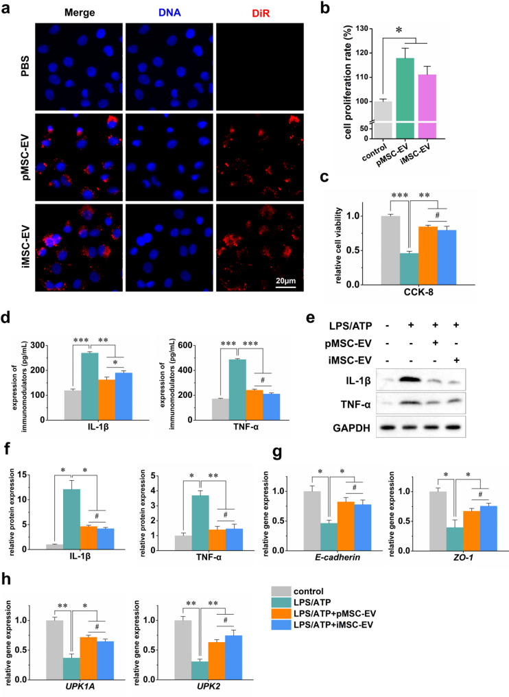
Methods: To overcome these challenges, we developed conditionally immortalized mesenchymal stem cells (iMSCs) using a doxycycline-regulated simian virus 40 Large T expression system in pMSCs. The conditioned proliferation capacity of iMSCs was evaluated using cell counting and immunocytochemistry. The expression of surface markers on iMSCs was assessed by flow cytometry. The osteogenic and adipogenic differentiation capabilities of iMSCs at different population doubling (PD) numbers were analyzed by qPCR, alizarin red staining, and oil red O staining. The EVs secreted by iMSCs were characterized using Western blot, scanning electron microscopy, and particle size analysis. In vitro and in vivo bladder inflammation models were used to evaluate the therapeutic effects of EVs on IC/BPS.

Results: These iMSCs exhibited precisely controlled proliferation, maintained surface marker expression and differentiation capacities, comparable to pMSCs up to PD 40. The characteristics of iMSC-EVs are equivalent to those of pMSCs. Furthermore, in vitro cellular experiments demonstrate that iMSC-EVs provide protective effects against LPS/ATP-induced damage in SV-HUC-1 cells. Additionally, administration of iMSC-EVs significantly enhanced tissue healing and anti-inflammatory capabilities in an IC/BPS animal model.

Conclusions: In summary, this approach produced a reliable source of functional MSCs and EVs, with iMSC-EVs demonstrating robust immunomodulatory properties and promoting tissue healing in IC/BPS. This method represents a promising alternative to pMSCs for IC/BPS therapy.

Keywords: Anti-inflammatory; Conditional immortalization; EVs; IC/BPS; MSCs.


Conflict of interest statement

Declarations. Ethics approval and consent to participate: (1) Title of the approved project: Conditional Immortalization of Human Mesenchymal Stem Cells and Therapeutic Potential of Their Extracellular Vesicles Against Interstitial Cystitis; (2) Name of the institutional approval committee: The Animal Ethics Committee of Longgang District People’s Hospital of Shenzhen. (3) Approval number: 2024083DW. (4) Date of approval: 30/October/2024. The original source (Cyagen) of MSCs has confirmed that there was initial ethical approval for collection of human cells, and that the donors had signed informed consent. The original source (Pricella) of SV-HUC-1 cells has confirmed that there was initial ethical approval for collection of human cells, and that the donors had signed informed consent. Consent for publication: All authors agree to publish this version of the article. Competing interests: The authors declare that they have no competing interests.

Figures

Fig. 1
Fig. 1
Generation of Conditionally Immortalized Human MSCs. (a) Schematic representation of the MSCs immortalization procedure. (b, c) Immunostaining (b) and Western blot analyses (c) confirming LT expression in transduced MSCs. Hoechst 33342 (blue) was used to stain cell nuclei. Full-length blots are presented in Supplementary Fig. 1. (d) Growth curves of pMSCs and transduced MSCs when cultured with dox. (e) Diagrammatic representation of the iMSC preparation process. (f, g) Western blot analyses (f) and immunostaining (g) of LT expression in iMSCs cultured with dox and after removal of dox at 2, 4, 6, and 8-day intervals. Cell nuclei were stained using Hoechst 33342 (blue). Full-length blots are presented in Supplementary Fig. 1. (h) Quantitative analysis of iMSC numbers in cultures with or without dox. MSCs: Mesenchymal stromal cells; LT: Simian virus 40 Large T; GAPDH: Glyceraldehyde-3-phosphate dehydrogenase; pMSCs: Primary MSCs; iMSCs: Conditionally immortalized MSCs; dox: Doxycycline. Data are presented as mean ± SD (n = 3)
Fig. 2
Fig. 2
Assessment of MSC-Specific Markers. Flow cytometry analysis of pMSCs and iMSCs under both dox-free and dox-containing conditions, assessing well-established MSC markers. Positive markers include CD105, CD73, and CD90, while negative markers include CD14, CD34, and CD45. MSCs: Mesenchymal stromal cells; pMSCs: Primary MSCs; iMSCs: Conditionally immortalized MSCs; dox: Doxycycline; iMSC + dox: Conditionally immortalized MSCs treated with doxycycline. Data are presented as mean ± SD (n = 3)
Fig. 3
Fig. 3
Osteogenic and Adipogenic Differentiation Capacities of iMSCs. (a, b) ALP activity as an indicator of early osteogenic differentiation. (c) RT-qPCR analysis of osteogenic markers, including RUNX2, OSX, OPN, and OCN. (d, e) Oil Red O staining revealing intracellular lipid accumulation following adipogenic induction. (f) RT-qPCR analysis of adipogenic markers, including PPARG, CEBPA, LPL, and FABP4. pMSCs: Primary MSCs; iMSCs: Conditionally immortalized MSCs; dox: Doxycycline; iMSC + dox: Conditionally immortalized MSCs treated with doxycycline; ALP: Alkaline phosphatase; RUNX2: Runt-related transcription factor 2; OSX: Osterix; OPN: Osteopontin; OCN: Osteocalcin; PPARG: Peroxisome proliferator activated receptor γ; CEBPA: CCAAT enhancer binding protein alpha Gene; LPL: Low density lipoprotein; FABP4: Fatty acid binding protein 4. Data are presented as mean ± SD (n = 3). *P < 0.05, **P < 0.01, and ***P < 0.001
Fig. 4
Fig. 4
Switching of iMSCs Between Immortalized and Non-Immortalized State. (a) Diagrammatic representation of the experimental design. (b) Immunostaining of LT and Ki-67 expression in iMSCs at the indicated time points. (c) ALP and ARS following osteogenic induction of iMSCs. (d, e) RT-qPCR analysis of osteogenic marker, including RUNX2, OSX, OPN, and OCN. iMSCs: Conditionally immortalized MSCs; LT: Simian virus 40 Large T; ALP: Alkaline phosphatase; ARS: Alizarin red staining; RUNX2: Runt-related transcription factor 2; OSX: Osterix; OPN: Osteopontin; OCN: Osteocalcin. Data are presented as mean ± SD (n = 3). **P < 0.01 and ***P < 0.001
Fig. 5
Fig. 5
Characterizing EVs Secreted from iMSCs. (a) Western blot analysis of EV markers. Full-length blots are presented in Supplementary Fig. 1. (b) Histograms of EV size determined using the NanoSight instrument. (c) Electron microscopy images of isolated EVs. (d) RT-qPCR analysis of miRNA-9, miRNA-146a-5p, and miRNA-223-5p expression in EVs. (e) ELISA analysis of immunoregulatory factors IL-10, PD-L1, and TGF-β1 levels in EVs. EVs: Extracellular vesicles; pMSCs: Primary MSCs; iMSCs: Conditionally immortalized MSCs; pMSC-EV: Primary MSCs-derived extracellular vesicles; iMSC-EV: Conditional immortalized MSCs-derived extracellular vesicles; IL-10: Interleukin-10; PD-L1: Programmed cell death ligand 1; TGF-β1: Transforming growth factor-β1. Data are presented as mean ± SD (n = 3). #P > 0.05
Fig. 6
Fig. 6
Protective Effects of iMSC-EVs on LPS/ATP-Induced Damage in SV-HUC-1 Cells. (a) Confocal microscopy images of SV-HUC-1 cells treated with DiR-labeled EVs. (b, c) Cell viability was assessed using the CCK-8 assay. (d-f) ELISA (d) and Western blot (e, f) measurement of inflammatory factors, including IL-1β and TNF-α. Full-length blots are presented in Supplementary Fig. 1. (f, g) RT-qPCR analysis of gene expression in SV-HUC-1 cells, including epithelial cell markers (E-cadherin and ZO-1) (f) and urothelial markers (UPK1A and UPK2) (g). pMSC-EV: Primary MSCs-derived extracellular vesicles; iMSC-EV: Conditional immortalized MSCs-derived extracellular vesicles; LPS: Lipopolysaccharide; ATP: Adenosine triphosphate; DiR: 1,1-dioctadecyl-3,3,3,3-tetramethylindotricarbocyaine iodide; IL-1β: Interleukin-1β; TNF-α: Tumor necrosis factor-α; ZO-1: Zona Occludens 1; UPK1A: Uroplakin-1 A; UPK2: Uroplakin-2. Data are presented as mean ± SD (n = 3). #P > 0.05, *P < 0.05, **P < 0.01, and ***P < 0.001
Fig. 7
Fig. 7
Therapeutic Activity of iMSC-EVs in an IC/BPS Mouse Model. (a) Experimental protocol for comparing the therapeutic effects of iMSC-EVs or pMSC-EVs in a CYP-induced IC/BPS mouse model. (b) Wet bladder weight measurement. (c) Histological examination of bladder morphology with H&E and MT staining. (d) Quantification of fibrotic areas from MT staining. (e-g) RT-qPCR (e) and Western blot (f, g) analysis inflammatory factors (Il-1b and Tnf-a) expression in bladder tissue. (h, i) RT-qPCR analysis of gene expression in bladder tissue, including urothelial markers (Upk1a and Upk2) (h) and IC/BPS-related biomarkers (Itgal and Psmb9) (i). (j) ELISA measurement of PGE2 and NAG levels in urine. CYP: Cyclophosphamide; EV: Extracellular vesicle; IC/BPS: Interstitial cystitis/bladder pain syndrome; pMSC-EV: Primary MSCs-derived extracellular vesicles; iMSC-EV: Conditional immortalized MSCs-derived extracellular vesicles; H&E: Hematoxylin and eosin; MT: Masson’s trichrome; Il-1b: Interleukin-1b; Tnf-a: Tumor necrosis factor-a; Upk1a: Uroplakin-1a; Upk2: Uroplakin-2; Itgal: Integrin subunit alpha l; Psmb9: Proteasome 20 S Subunit Beta 9; PGE2: Prostaglandin E2; NAG: N-acetyl-β-D-glucosaminidase; Data are presented as mean ± SD (n = 3). #P > 0.05, *P < 0.05, **P < 0.01, and ***P < 0.001

References

    1. C MCHA, et al. Assessment of treatment outcomes of interstitial cystitis with hydrodistention and bladder training by O’Leary-Sant interstitial cystitis symptom and problem indices. Taiwan J Obstet Gynecol. 2018;57(5):718–21. - PubMed
    1. Homma Y et al. Clinical guidelines for interstitial cystitis/bladder pain syndrome. International Journal of Urology, 2020(Suppl 1).
    1. A KJK, et al. Comparison of the efficacy between transurethral coagulation and transurethral resection of Hunner lesion in interstitial cystitis/bladder pain syndrome patients: A prospective randomized controlled trial. Eur Urol. 2020;77(5):644–51. - PubMed
    1. Hanno PM, et al. Diagnosis and treatment of interstitial cystitis/bladder pain syndrome: AUA guideline amendment. J Urol. 2015;193(5):1545–53. - PubMed
    1. Mullins C, et al. Novel research approaches for interstitial cystitis/bladder pain syndrome: thinking beyond the bladder. Translational Androl Urol. 2015;4(5):524–33. - PMC - PubMed

LinkOut - more resources