. 2025 Aug 26;16(1):15.
doi: 10.1186/s13227-025-00251-9.

Predetermined sex revealed by a female transient gut in non-feeding larvae of Osedax (Siboglinidae, Annelida)

Affiliations

Predetermined sex revealed by a female transient gut in non-feeding larvae of Osedax (Siboglinidae, Annelida)

Alice Rouan et al. Evodevo. .

Abstract

Background: Within the symbiont-hosting Siboglinidae (Annelida), Osedax stands out as the sole genus capable of degrading bones and displaying pronounced sexual dimorphism (except O. priapus). While macroscopic, gutless females feed on whale falls with their symbiont-housing "roots", males are microscopic and non-feeding. Yet, embryos and larvae look identical, and sex is suggested to be environmentally determined, i.e., larvae metamorphose into females on bare bone or into males when finding an adult female.

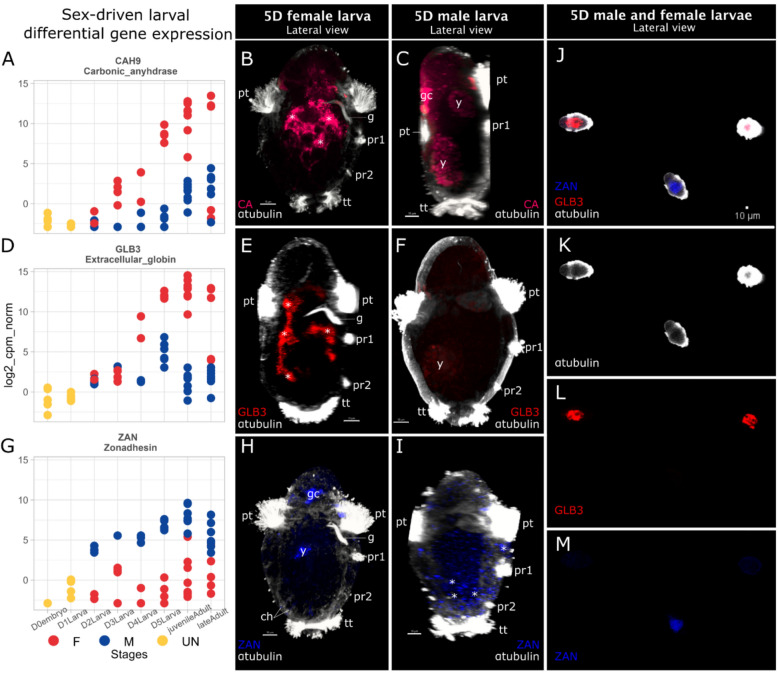
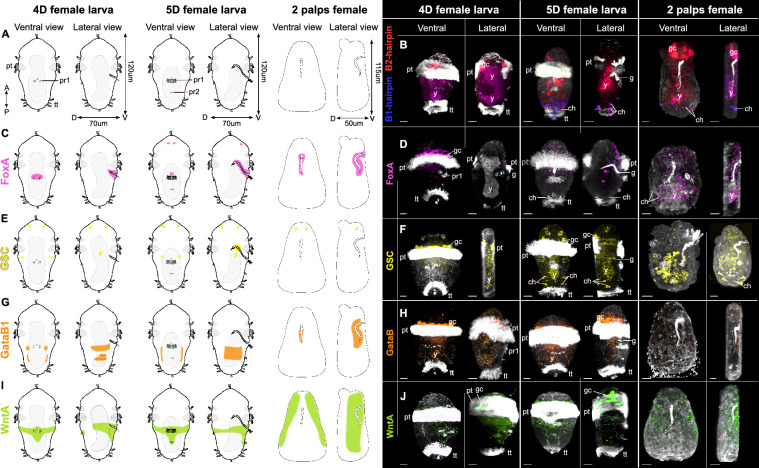
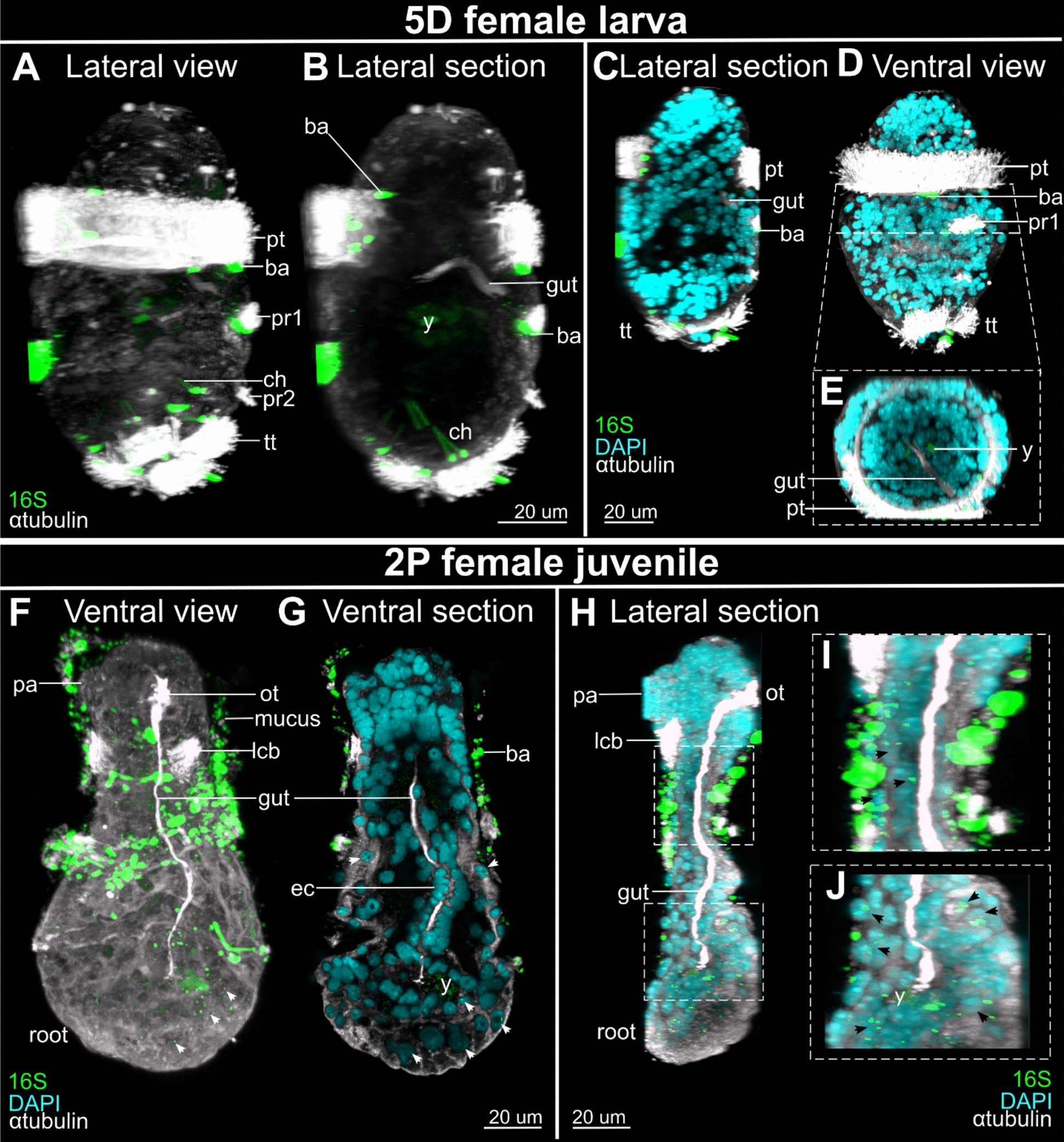
Results: However, we here describe a transient gut present in half of the late larvae and in juvenile females of O. japonicus. We confirm the gut-carrying larvae as being females from sex-specific in situ gene expression. Moreover, morphological evidence coupled with differential gene expression indicate that the 'non-feeding' transient gut may pattern the vascular system and/or act as a gas-exchange surface in juvenile females, before their branchial appendages develop.

Conclusions: The transient gut of O. japonicus females reveals a genetic sex determination. Proposedly homologous across siboglinids, this vestigial gut is suggested to function in organ patterning and/or for gas-exchange during development of the gutless adult.

Keywords: Lecithotrophic larvae; Rudimentary gut; Sex determination; Sexual dimorphism; Trochophore; Vascular system.


Conflict of interest statement

Declarations. Ethics approval and consent to participate: Not applicable. Consent for publication: Not applicable. Competing interests: The authors declare no competing interests.

Figures

Fig. 1
Fig. 1
Osedax japonicus gut development from the 4-day-old (4D) larva to the 2 palps (2P) juvenile female stage. A Confocal laser scanning microscopy (CLSM) of anti-acetylated α-tubulin (white) stained cilia showing lateral views of 4D and 5D larvae, early metamorphosed females, and early 2P females, ventral views of later 2P stages and mature adult 4P stage. Lateral projections were virtually sectioned to show the gut in larval stages, B scaled corresponding drawings in lateral view (dorsal to the left, ventral to the right); the 4P drawing is reduced 2.5× in size. ao apical organ, cc circumesophageal connectives, D–V dorso-ventral, do dorsal organ, dpi days post-induction, g gut, hpc hours post-cleavage, hpi hours post-induction, lcb lateral ciliary band, m mouth, ot oral tuft, P/pa palp, pi pinnules, pr1-2 paratroch 1–2, pt prototroch, tert terminal tuft, tt telotroch. Scale bars: 20 µm
Fig. 2
Fig. 2
Differential gene expression between sexes in larval stages of O. japonicus. ADG Log2 scaled normalized gene expression (cpm) of carbonic anhydrase (CAH9), extracellular globin (GLB3) and zonadhesin (ZAN). Individual dots show individual samples of undetermined sex (yellow), female (red), male (blue), B, CE, FH, I dorso-ventral (left to right) view of maximum intensity z-projection of whole mount in situ-HCR CLSM scans showing CAH9 (dark pink), GLB3 (red) and ZAN (blue) coupled with acetylated α-tubulin-like immunoreactivity (white) in female (left) and male (right) 5-days-old (5D) larvae. Lateral projections were virtually sectioned to remove prototroch signals for better display of intended structures. J WMIS–HCR CLSM scans of 5D larvae siblings showing K anti-acetylated α-tubulin-like immunoreactivity (white) L GLB3 (red) expression and M ZAN (blue) expression. 5D 5-day-old, ch chaetae, g gut, gc glandular cell, pr1-2 paratroch 1–2, y yolk. Asterisks (*) highlight signals. Scale bars: 10 µm
Fig. 3
Fig. 3
Ventral and lateral views of temporal expression pattern visualized by whole mount in situ-HCR coupled with anti-acetylated α-tubulin immunostaining (white) in 4-day-old (4D), 5D female larvae and early 2 palps (2P) females. A Scaled drawings of female ventral and leral view, B HCR negative control using only B1-(blue) and B2-(red) hairpins highlighting the yolk, pigmented prototroch cells and chaetae background, C, D drawing and HCR of FoxA (pink) expression pattern, E, F drawing and HCR of Goosecoid (GSC) (yellow) expression pattern, G, H drawing and HCR of GataB1 (orange) expression pattern, I, J drawing and HCR of WntA (green) expression pattern. CEGI Drawings represent the conserved signal found across individuals of the same stages. Blue dotted lines highlight the probes’ signal. Lateral projections were virtually sectioned to remove prototroch signals for better display of intended structures in lateral views. 4D/5D 4/5-day-old, ch chaetae, D dorsal, gc glandular cell, pt prototroch, pr1/2 paratroch 1/2, tt telotroch, V ventral, y yolk. Scale bars: 15 µm
Fig. 4
Fig. 4
Ventral views and lateral views (ventral to the right) of symbiont localization by whole mount in situ-HCR in O. japonicus 5-days-old (5D) female larvae (A-E) and 2-palps (2P) females (F-J). A Lateral, maximum z-projection only showing bacterial aggregates externally among cilia, using 16S probes (green). B Mid-sagittal view, achieved by virtually removing outermost lateral sections, showing lack of bacterial signal in or near the gut. C Mid-sagittal view, showing nuclei signal (DAPI, cyan). D Ventral, maximum z-projection only showing external bacterial aggregates. E Cross section of gut region showing no bacterial signal in the endoderm gut cells. F Ventral, maximum z-projection with bacterial 16S probe signal externally, trapped in mucus, and internally, within cells of the root and trunk (arrowheads).G Sub-ventral view, achieved by virtually removing outermost ventral sections, showing lack of bacterial signal in gut. H Mid-sagittal view of multiple sections, showing similar signal as F. I-J, close-up of H showing external bacterial aggregates in mucus and internal signal in lower trunk (I) and root (J) tissue. ba bacteria aggregates, ch chaetae, ec endoderm gut cell, lcb lateral ciliary band, ot oral tuft, pa palp, pr1-2 paratroch 1–2, pt prototroch, tt telotroch, y yolk. Arrowhead highlight signals. Scale bars 20 µm
Fig. 5
Fig. 5
Ventro-lateral views of O. japonicus CLSM phalloidin stain (red–yellow gradient) showing the heart development relative to the gut (anti-acetylated α-tubulin stain, white) and nuclei (DAPI, cyan). A 5-days-old (D) larva showing the position of the gut relative to the musculature (red–yellow gradient). B Mid-anterior trans-section view. C Dorso-ventral (left–right) longitudinal section showing actin staining near the gut. D Early metamorphosed female showing the elongation of the gut medioventrally. E Medioventral trans-section showing the heart and gut proximity. F Dorso-ventral (left–right) longitudinal section showing the cellular contact between heart and gut. G 2-palps (2P) female stage showing the longitudinal elongation of the gut with the trunk and its direct contact with the heart body. H, I Enlargement of the heart body. C, DF, G, H, I Lateral stacks were virtually removed from maximal z-projection to show the heart structure and removed longitudinal muscle. Abbreviations: dc degenerated cilia, g gut, h heart, lcb lateral ciliary band, lm longitudinal muscle, pa palp, pr1-2 paratroch 1–2, pt prototroch, tt telotroch. Scale bars: AG 10 µm and H, I 7 µm
Fig. 6
Fig. 6
New life cycle hypothesis of the bone-eating worm Osedax having sexually predetermined larvae. Schematic drawing of Osedax development with sexually determined embryos developing into sexually determined larvae, female larvae having a gut, male larvae lacking one. Drawings are not scaled

References

    1. Cavanaugh CM, Gardiner SL, Jones ML, Jannasch HW, Waterbury JB. Prokaryotic cells in the hydrothermal vent tube worm Riftia pachyptila Jones: possible chemoautotrophic symbionts. Science. 1981;213(4505):340–2. - PubMed
    1. Rouse GW, Goffredi SK, Vrijenhoek RC. Osedax : bone-eating marine worms with dwarf males. Science. 2004;305(5684):668–71. - PubMed
    1. Southward EC, Schulze A, Gardiner SL. Pogonophora (Annelida): form and function. Hydrobiologia. 2005;535(536):227–51.
    1. Berman G, Johnson S, Seid C, Vrijenhoek R, Rouse G. Range extensions of Pacific bone-eating worms (Annelida, Siboglinidae, Osedax). BDJ. 2023;11: e102803. - PMC - PubMed
    1. Goffredi SK, Orphan VJ, Rouse GW, Jahnke L, Embaye T, Turk K, Lee R, Vrijenhoek RC. Evolutionary innovation: a bone-eating marine symbiosis. Environ Microbiol. 2005;7:1369–78. - PubMed

LinkOut - more resources