. 2025 Jul 25;21(11):4834-4850.
doi: 10.7150/ijbs.105629. eCollection 2025.

Methylation-mediated LncRNA CRAT40 promotes colorectal cancer progression by recruiting YBX1 to initiate RelA transcription

Affiliations

Methylation-mediated LncRNA CRAT40 promotes colorectal cancer progression by recruiting YBX1 to initiate RelA transcription

Qing Lu et al. Int J Biol Sci. .

Abstract

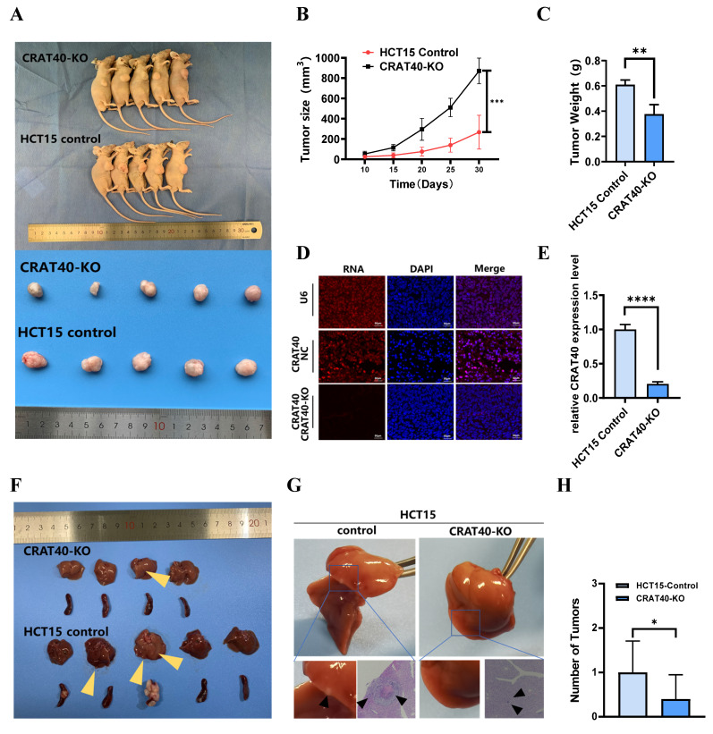
Colorectal cancer (CRC) remains a leading cause of cancer-related mortality worldwide. Long noncoding RNAs (lncRNAs) have emerged as crucial regulators in the initiation and progression of various malignancies, including CRC. In this study, we found that lnc-CRAT40 was upregulated in CRC and associated with poor prognosis following CRC resection. Functional assays revealed that elevated lnc-CRAT40 expression promotes tumor cell proliferation and metastasis both in vitro and in vivo. The modification of N6-methyladenosine, driven by METTL3, was essential for the stability of lnc-CRAT40, which may partially contribute to the upregulation of lnc-CRAT40. Mechanistically, lnc-CRAT40 directly interacted with Y-box binding protein 1 (YBX1) and recruits it to the RelA promoter, thereby activating NF-κB signaling, which in turn drives CRC proliferation and metastatic potential. These findings provide novel insights into the molecular mechanisms underlying CRC progression and highlight lnc-CRAT40 as a potential prognostic biomarker and therapeutic target.

Keywords: METTL3; YBX1; colorectal cancer; long non-coding RNA.


Conflict of interest statement

Competing Interests: The authors have declared that no competing interest exists.

Figures

Figure 1
Figure 1
LncRNA CRAT40 is upregulated in CRC and associated with poor prognosis. A. Volcano plot of RNA-seq results showing differentially expressed lncRNAs in 5 pairs of CRC tumor and adjacent normal tissues. Each dot represents a gene; red dots indicate significantly upregulated lncRNAs, green dots indicate significantly downregulated ones. B. Heatmap of selected differentially expressed lncRNAs, with lnc-CRAT40 highlighted in red. C. Quantitative analysis of lnc-CRAT40 expression in 103 paired CRC and adjacent normal tissues by qRT-PCR. D. Kaplan-Meier survival analysis of overall survival in CRC patients stratified by lnc-CRAT40 expression levels (log-rank test). E. Expression of lnc-CRAT40 in unpaired CRC and normal tissues from the TCGA dataset. F. lnc-CRAT40 expression levels in normal colon epithelial cells (HCoEpiC) and CRC cell lines. Data are presented as mean ± SD. N, normal tissues; T, tumor tissues. **P<0.01, ***P<0.001, ****P<0.0001.
Figure 2
Figure 2
Silencing of Lnc-CRAT40 inhibits CRC proliferation, migration and invasion in vitro. A. Schematic of CRISPR/Cas9-mediated knockout of lnc-CRAT40 in HCT15 cells using paired sgRNAs targeting sequences flanking the transcriptional locus, resulting in a 1.1 kb deletion. B. Identification of knockout clones by genomic PCR and confirmation of homozygous deletion by Sanger sequencing; off-target regions served as negative controls. C. qRT-PCR analysis of lnc-CRAT40 expression in SW480 and HCT15 cells after CRAT40 silencing or control transfection (left), and in control versus CRAT40 knockout HCT15 clones (right). D. Cell proliferation assessed by CCK-8 assay following lnc-CRAT40 knockdown or knockout. E, F. Wound healing and Transwell assays evaluating migration and invasion in knockdown or knockout cells compared with controls; quantification shown on right. Data represent mean ± SD from three independent experiments. NC, negative control; KO, knockout. *P<0.1, **P<0.01, ***P<0.001, ****p<0.0001.
Figure 3
Figure 3
Knockout of lnc-CRAT40 inhibits CRC proliferation, migration and invasion in vivo. A. Representative images of xenograft tumors from control and CRAT40 KO groups. B, C. Quantification of tumor weights and volumes. Data represent mean ± SD from five independent mice per group. D. FISH images showing lnc-CRAT40 signal in tumor tissues from control and KO groups; scale bar, 50 μm. E. qRT-PCR detection of lnc-CRAT40 expression in xenograft tumors. F. Representative images of liver metastatic nodules following intrasplenic injection. G. H&E staining of liver metastatic lesions. H. Quantification of metastatic nodules in liver tissue. Data shown as mean ± SD. H&E hematoxylin and eosin; NC, negative control; KO, knockout. *P<0.05, **P<0.01, ***P<0.001, ****P<0.0001.
Figure 4
Figure 4
Lnc-CRAT40 regulates RelA expression in CRC cells. A. Subcellular fractionation revealing nuclear localization of lnc-CRAT40 in CRC cell lines. B. Representative FISH images of lnc-CRAT40 localization in SW480 and HCT15 cells (red); nuclei stained with DAPI (blue). Scale bar, 20 μm. C. Heatmap showing top 15 upregulated and downregulated genes after CRAT40 knockout in HCT15 cells. D, E. qRT-PCR and Western blot analyses of RelA mRNA and p65 protein levels following lnc-CRAT40 knockdown or knockout. F. Upregulation of RelA in TCGA CRC cohort. G. qRT-PCR validation of RelA expression in 72 paired CRC tissues and adjacent noncancerous tissues. H. Correlation analysis between lnc-CRAT40 and RelA expression in CRC samples (GEPIA 2 database). I. ChIRP assay showing enrichment of lnc-CRAT40 at the RelA promoter region in HCT15 cells. COAD, colon adenocarcinoma; READ, rectum adenocarcinoma; N, normal tissues; T, tumor tissues; NC, negative control; ChIRP, chromatin isolation by RNA; ns, not significant. *P<0.05, **P<0.01, ***P<0.001, ****P<0.0001.
Figure 5
Figure 5
Lnc-CRAT40 interacts with YBX1. A, B. Silver staining of proteins pulled down from SW480 cells using biotin-labeled NC or lnc-CRAT40. The arrow indicates the band corresponding to YBX1, identified by mass spectrometry. C. Secondary mass spectrogram of YBX1 peptide identified in pull-down. D. Western blot confirming specific binding between YBX1 and lnc-CRAT40 in RNA pull-down assays. E. Western blot validating enrichment of YBX1 antibody in RIP assays compared with IgG control. F. RIP-qPCR showing enrichment of lnc-CRAT40 in YBX1 immunoprecipitates from SW480 and HCT15 cells. G. RNA pull-down using wild-type and mutant lnc-CRAT40 probes assessed for YBX1 binding. H. Co-localization of lnc-CRAT40 (FISH) and YBX1 (immunofluorescence) in CRC cells. I, J. Proliferation, migration, and invasion assays showing that YBX1 knockdown reverses phenotypes induced by lnc-CRAT40 overexpression. Data are presented as means ± SD and were analyzed using Student's t-test; all experiments were performed in triplicate. NC, negative control; KD, knockdown; Ctrl, control; mut, mutant; IP, immunoprecipitation. *P<0.1, **P<0.01.
Figure 6
Figure 6
Lnc-CRAT40 recruits YBX1 to co-activate RelA transcription. A, B. Genome-wide distribution of YBX1-binding sites in HCT15 cells identified by ChIP-seq, with 60.7% located at promoter regions. C. Motif analysis of YBX1 binding sequences. D. ChIP-seq tracks showing YBX1 peaks at the RelA promoter. E. Schematic of PCR-amplified regions within the RelA promoter for ChIP-qPCR. F. ChIP-qPCR confirming specific YBX1 binding to the P2 region of the RelA promoter; IgG as negative control. G, H. ChIP-qPCR showing reduced YBX1 enrichment at the RelA promoter after YBX1 knockdown or lnc-CRAT40 knockout. I. Western blot showing decreased p65 protein after YBX1 knockdown. J, K. Functional rescue assays: RelA knockdown inhibits proliferation, migration, and invasion, which are reversed by YBX1 overexpression. L. Western blot analysis of p65 following YBX1 overexpression in RelA-silenced cells. M. Western blot of NF/κB and EMT pathway-related proteins, including Bcl-2 and E-cadherin, after YBX1 knockdown and lnc-CRAT40 knockout. ChIP-seq, chromatin immunoprecipitation sequencing; KD, knockdown; NC, negative control; TSS, transcription start site; ns, not significant. *P<0.1, **P<0.01, ***P<0.001.
Figure 7
Figure 7
Lnc-CRAT40 is upregulated in CRC via METTL3-mediated m6A modification. A. Predicted m6A modification sites within lnc-CRAT40 transcript (SRAMP database). B. The decrease of lnc-CRAT40 expression in HCT15 and SW480 cells treated with methylation inhibitor 3-DAA for 24 h (qRT-PCR). C. Quantitative analysis of METTL3 expression in 49 paired CRC and adjacent normal tissues by qRT-PCR. D. Elevated METTL3 expression in CRC tissues (TCGA dataset). E. Correlation between lnc-CRAT40 and METTL3 expression in CRC tissue (n=49). F. Positive correlation between lnc-CRAT40 and METTL3 expression in CRC (GEPIA 2). G. Kaplan-Meier analysis showing worse disease-free survival in CRC patients with high METTL3 expression. H. Western blot confirming METTL3 knockdown efficiency in CRC cells. I. qRT-PCR showing reduced METTL3 and lnc-CRAT40 expression upon METTL3 knockdown. J. MeRIP-qPCR demonstrating decreased m6A modification on lnc-CRAT40 following METTL3 knockdown. K, L. RNA stability assay showing shortened half-life of lnc-CRAT40 transcript after METTL3 knockdown. Data are mean ± SD of three independent experiments. *P < 0.1, **P < 0.01, ***P < 0.001, ****P < 0.0001.
Figure 8
Figure 8
Schematic model illustrating the mechanism of lnc-CRAT40 in CRC progression. Schematic overview depicting that METTL3-mediated m6A modification stabilizes lnc-CRAT40, which functions as a scaffold recruiting YBX1 to the RelA promoter, activating NF-κB signaling and promoting colorectal cancer proliferation and metastasis.

Similar articles

References

    1. Global Burden of Disease 2019 Cancer Collaboration; Kocarnik JM, Compton K. et al. Cancer Incidence, Mortality, Years of Life Lost, Years Lived With Disability, and Disability-Adjusted Life Years for 29 Cancer Groups From 2010 to 2019: A Systematic Analysis for the Global Burden of Disease Study 2019. JAMA Oncol. 2022;8:420–444. - PMC - PubMed
    1. Biller LH, Schrag D. Diagnosis and Treatment of Metastatic Colorectal Cancer: A Review. JAMA. 2021;325:669–685. - PubMed
    1. Adnane S, Marino A, Leucci E. LncRNAs in human cancers: signal from noise. Trends Cell Biol. 2022;32:565–573. - PubMed
    1. Chang L, Ding J, Pu J. et al. A novel lncRNA LOC101928222 promotes colorectal cancer angiogenesis by stabilizing HMGCS2 mRNA and increasing cholesterol synthesis. J Exp Clin Cancer Res. 2024;43:185. - PMC - PubMed
    1. Liu H, Li D, Sun L. et al. Interaction of lncRNA MIR100HG with hnRNPA2B1 facilitates m6A-dependent stabilization of TCF7L2 mRNA and colorectal cancer progression. Mol Cancer. 2022;21:74. - PMC - PubMed

MeSH terms