. 2025 Aug 21:14:849-875.
doi: 10.2147/ITT.S533552. eCollection 2025.

Fracture-Induced Immunological Cascades Trigger Rapid Systemic Bone Loss via Osteocyte-Regulated Osteoclastogenesis

Affiliations

Fracture-Induced Immunological Cascades Trigger Rapid Systemic Bone Loss via Osteocyte-Regulated Osteoclastogenesis

Lipeng Sun et al. Immunotargets Ther. .

Abstract

Background: Rapid bone loss after fracture elevates the risk of subsequent fractures, but the mechanisms remain unclear. IL-6, a key cytokine involved in fracture healing, is markedly upregulated during the immune response after fracture; however, its role in systemic skeletal deterioration remains poorly defined.

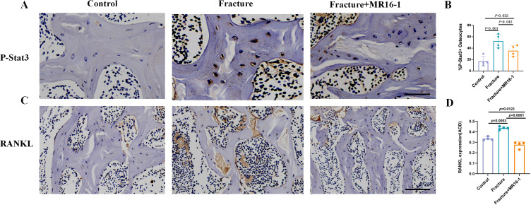
Methods: In this study, we employed label-free proteomics to identify candidate mediators in vertebral samples following fracture. Next, osteocyte siRNA knockdown and Stattic (STAT3 phosphorylation inhibitor) inhibition were used to investigate IL-6 related signaling pathways. Subsequently, indirect co-cultures of osteocyte with osteoclast or osteoblast were used to evaluate the effects of the IL-6 pathway on bone resorption and formation. Furthermore, fractured mice were treated with MR16-1 (monoclonal anti-mouse IL-6 receptor antibody) or Stattic. Then, trabecular and cortical bone in vertebrae and femur were evaluated at 4, 14, and 28 days post-fracture, including histological analysis of p-STAT3+ osteocyte, RANKL expression, and bone formation/resorption markers.

Results : In vitro, IL-6 dose-dependently elevated RANKL and p-STAT3 levels in osteocyte and promoted osteoclast activity in co-culture. These effects were suppressed by Stattic and replicated by STAT3 knockdown. In contrast, co-culture of osteocyte with osteoblast exhibited no significant alterations in osteogenic marker expression upon IL-6 exposure, suggesting negligible effects on osteoblast activity. In vivo, MR16-1 reduced trabecular bone loss in the vertebrae and femur after fracture. It also diminished p-STAT3+ osteocyte, reduced RANKL expression, and suppressed osteoclast activity without impairing osteoblastogenesis. And Stattic produced a comparable reduction in systemic bone loss and osteoclast overactivation.

Conclusion: This study demonstrates that IL-6 drives osteoclast-mediated bone resorption via STAT3-dependent RANKL induction in osteocyte, thereby aggravating post-fracture systemic bone loss. And the findings highlight that modulating the IL-6/STAT3/RANKL axis and targeting osteocyte function may offer a promising therapeutic approach for preventing bone loss and minimizing the risk of fracture recurrence.

Keywords: RANKL; fracture healing; fracture risk; inflammation; osteocyte; osteoporosis.


Conflict of interest statement

The authors declare that they have no competing interests in this work.

Figures

Figure 1
Figure 1
Animal treatment protocol.The initial red arrow indicates that fracture surgery was performed on day 0, followed immediately by the first MR16-1 or Stattic injection. The second red arrow represents the administration of Sequential fluorescence labeling, including calcein green and alizarin red, for dynamic bone formation assessment. Black arrows denote time points for sample collection and analyses, including micro-CT, TRAP staining, inflammatory cytokine quantification, immunohistochemistry, and immunofluorescence. These evaluations were conducted at days 4, 14, and 28 post-fracture.
Figure 2
Figure 2
IL-6 levels rapidly increase both locally and systemically after femur fracture, and the use of MR16-1 blocks this increase. (A) KEGG enrichment analysis. Differentially expressed proteins were defined by t-test (p < 0.05, fold change >1.2 or <0.83); “***” indicates p < 0.001 for pathway enrichment significance. (n = 2 mice/group). (B) Protein interaction analysis. (C) Representative images showing the localization of IL-6 fluorescence at the fracture sites in three groups: saline-injected mice, MR16-1-injected mice, and control mice. The scale bar represents 250µm. (D) Serum IL-6 levels measured at 4 days, 2 weeks, and 4 weeks after fracture in mice treated with saline, MR16-1, and control (n = 5 mice/group). (E) Serum TNF-α levels measured at 4 days, 2 weeks, and 4 weeks after fracture in mice treated with saline, MR16-1, and control (n = 5 mice/group). (F) Serum IL-1β levels measured at 4 days, 2 weeks, and 4 weeks after fracture in mice treated with saline, MR16-1, and control (n = 5 mice/group). Error bars indicate standard deviations (SDs). p-values were calculated using one-way ANOVA followed by Tukey’s post hoc test.
Figure 3
Figure 3
Rapid cancellous bone loss in the L5 vertebra occurs after femur fracture, and the use of MR16-1 alleviates bone loss.(AI) Representative images of the 3D trabecular structure in the L5 vertebral body at 4 days, 2 weeks, and 4 weeks post-fracture in mice treated with saline, MR16-1, or the control group. (JU) Quantitative analysis of trabecular microarchitecture changes in the L5 vertebral body at 4 days, 2 weeks, and 4 weeks post-fracture in mice treated with saline, MR16-1, or control (n = 5 per group). Error bars indicate standard deviations (SDs). p-values were calculated using one-way ANOVA followed by Tukey’s post hoc test.
Figure 4
Figure 4
Fracture leads to bone loss in the contralateral distal femur metaphasis, and the use of MR16-1 can prevent bone loss.(AI) Representative images showing the three-dimensional trabecular structure of the contralateral distal femoral metaphysis at 4 days, 2 weeks, and 4 weeks post-fracture in mice treated with saline, MR16-1, or the control group. (JU) Quantitative analysis of changes in the trabecular microarchitecture of the distal femoral metaphysis at 4 days, 2 weeks, and 4 weeks after fracture in saline treated, MR16-1 treated, and control mice (n = 5 mice/group). Error bars represent standard deviations (SDs). p-values were obtained using one-way ANOVA followed by Tukey’s post hoc test.
Figure 5
Figure 5
The cortical bone of the contralateral femoral midshaft is unaffected after femur fracture, and the use of MR16-1 does not result in further changes. (AI) Representative cross-sectional images of the contralateral femoral midshaft at 4 days, 2 weeks, and 4 weeks post-fracture in mice treated with saline, MR16-1, or the control group. (JX) Quantitative µCT measurements of cortical thickness (Cort.Th), total cross-sectional area (Tt.Ar), cortical area (Ct.Ar), cortical area/total cross-sectional area (Ct.Ar/Tt.Ar), and medullary area (Ma.Ar) at 4 days, 2 weeks, and 4 weeks post-fracture in mice treated with saline, MR16-1, or control (n = 5 per group). Error bars represent standard deviations (SDs). p-values were calculated using one-way ANOVA followed by Tukey’s post hoc test.
Figure 6
Figure 6
Osteoclast activity increases promptly after fracture, and the use of MR16-1 can counteract this effect. (AI) Representative TRAP staining images of trabecular bone within the L5 vertebral body from control mice and fractured mice treated with saline or MR16-1 at (AC) 4 days, (DF) 2 weeks, and (GI) 4 weeks post-fracture. Scale bar = 250μm. (J and K) Mice with femur fractures exhibited a significantly higher number of osteoclast and resorbed surfaces compared to control mice at 4 days post-fracture, and these effects were reversed by MR16-1 treatment (n = 5 mice/group). (L–O) No significant differences were found at 2 and 4 weeks after fracture (n = 5 mice/group). Error bars represent standard deviations (SDs). p-values were calculated using one-way ANOVA followed by Tukey’s post hoc test.
Figure 7
Figure 7
Osteoblast activity does not significantly change after fracture, whereas the increase in osteoclast activity can be reversed by MR16-1 or Stattic treatment.(A) CTSK staining in control, fracture, and MR16-1 treated mice. (B) Quantification of CTSK fluorescence intensity (n = 4 mice/group). (C) OCN staining in control, fracture, and MR16-1 treated mice. (D) Quantification of OCN fluorescence intensity (n = 4 mice/group). (E) CTSK staining in control, fracture, and Stattic treated mice. (F) Quantification of CTSK fluorescence intensity (n = 4 mice/group). (G) OCN staining in control, fracture, and Stattic treated mice. (H) Quantification of OCN fluorescence intensity (n = 4 mice/group). Nuclei were counterstained with DAPI (blue), and CTSK and OCN positive signals are shown in red. Scale bars: 250 µm. Error bars represent standard deviations (SDs). p-values were calculated using one-way ANOVA followed by Tukey’s post hoc test. (I) Sequential fluorescence labeling using calcein and alizarin red in the trabecular bone of the L5 vertebral body in fractured mice, fractured mice treated with MR16-1, and control mice. Scale bar = 50 μm. (J-L) Dynamic histomorphometric analysis of trabecular bone formation following fracture. (n = 4 mice/group). Error bars represent standard deviations (SDs). p-values were determined using one-way ANOVA followed by Tukey’s post hoc test.
Figure 8
Figure 8
P-STAT3+ positive osteocyte and RANKL expression also increase after fracture, and these changes are attenuated by the use of MR16-1.(A) Representative image of osteocyte positively stained for P-STAT3 protein. Scale bar = 50 µm. (B) The percentage of P-STAT3+ osteocyte in control mice, fractured mice and fracture with MR16-1 treatment mice (n = 4 mice/group). (C) Immunohistochemical staining of RANKL in the cancellous bone of the L5 vertebra. Scale bar = 200 µm. (D) Quantification of RANKL levels of (C). (n = 4 mice/group) Error bars represent standard deviations (SDs). p-values were determined using one-way ANOVA followed by Tukey’s post hoc test.
Figure 9
Figure 9
IL-6 can stimulate osteocyte to secrete RANKL and increase osteoclast formation in an indirect coculture system of osteocyte and osteoclast. (A) Schematic of the protocol for the indirect co-culture of osteocyte and osteoclast to investigate the role of IL-6. (B) Western blot analysis of RANKL expression in MLO-Y4 cells following IL-6 activation. (C) Quantification of RANKL protein levels from (B) (n = 3). (D) Western blot analysis of osteoclast-specific markers, including NFATC1 and CTSK, following indirect co-culture. (E and F) Quantification of NFATC1 and CTSK protein levels from (D) (n = 3). (G) Representative confocal microscopy images showing co-staining of p-STAT3 (red), RANKL (green), and DAPI (blue) in MLO-Y4 cells. Scale bars = 25 µm. (H-I) Quantitative analysis of p-STAT3 and RANKL fluorescence intensity in MLO-Y4 cells stimulated with 0, 10, or 50 ng/mL IL-6. Both p-STAT3 and RANKL levels increased in a dose-dependent manner following IL-6 treatment, with statistically significant differences observed between all groups. (n = 3). (G) TRAP staining to assess osteoclast differentiation. Scale bars = 200 μm. (K) Osteoclast area was quantified following treatment with conditioned media derived from MLO-Y4 cells stimulated with increasing concentrations (0, 10, or 50 ng/mL) of recombinant mouse IL-6, corresponding to CM1, CM2, and CM3, respectively (n = 3). Error bars represent±SDs. All data are representative of at least three independent experiments. Statistical analysis was performed using one-way ANOVA followed by Tukey’s post hoc test.
Figure 10
Figure 10
IL-6 stimulates osteocyte to release RANKL, which indirectly increases osteoclast activity and relies on the phosphorylation of STAT3.(A) Experimental setup for the indirect coculture of osteocyte and osteoclast to investigate the role of STAT3. (B) Western blot analysis showing the expression of RANKL and phosphorylated STAT3 (P-STAT3) in MLO-Y4 cells following IL-6 stimulation and Stattic treatment. (C and D) Quantification of P-STAT3 and RANKL protein levels from (B). (n = 3). (E) Western blot analysis of osteoclastic marker genes, NFATC1 and CTSK, after indirect coculture with Stattic. (F and G) Quantification of NFATC1 and CTSK protein levels from (E). (n = 3). (H) Representative confocal microscopy images displaying co-staining of P-STAT3 (red), RANKL (green), and DAPI (blue). Scale bars = 25 µm. (I and J) Quantitative fluorescence analysis of p-STAT3 and RANKL expression in MLO-Y4 cells treated with IL-6 (50 ng/mL) in the presence or absence of Stattic (5 μM). IL-6 stimulation markedly increased p-STAT3 and RANKL levels, whereas co-treatment with Stattic significantly reduced both signals (n = 3). (K) TRAP staining to assess osteoclast differentiation. Scale bars = 200 μm. (K) Quantification of osteoclast area from (I). (n = 3). CM1 and CM3 were conditioned media collected from MLO-Y4 cells treated with 0 and 50 ng/mL recombinant mouse IL-6, respectively. CM4 was derived from MLO-Y4 cells treated with 50 ng/mL recombinant mouse IL-6 in combination with 5 µM Stattic. Error bars represent ± SDs. Data are representative of at least three independent experiments and were analyzed using one-way ANOVA followed by Tukey’s post hoc test.
Figure 11
Figure 11
Osteocyte STAT3 knock-down attenuates IL-6-induced RANKL expression, whereas conditioned media from IL-6-activated osteocyte do not alter osteoblast activity. (A) Western blot confirming efficient STAT3 knockdown in MLO-Y4 cells. (B) Quantification of STAT3 protein levels (n = 3). Followed by an unpaired Student’s t-test. (C) Western blot analysis of p-STAT3 and RANKL in osteocyte treated with IL-6 (50 ng/mL), with or without STAT3 siRNA. (D and E) Quantification of p-STAT3 and RANKL signals (n = 3). (F) Western blot analysis of ALP, RUNX2, and OCN in MC3T3-E1 cells cultured with osteocyte-conditioned media (CM1, CM2, or CM3). CM1, CM2, and CM3 were derived from MLO-Y4 cells exposed to 0, 10, or 50 ng/mL IL-6, respectively. (GI) Quantification of ALP, RUNX2, and OCN expression in CM1-CM3 groups. (n = 3). (J) Western blot analysis of ALP, RUNX2, and OCN in MC3T3-E1 cells treated with CM1, CM3, or CM4. CM4 represents conditioned medium from osteocyte stimulated with 50 ng/mL recombinant mouse IL-6 and 5 µM Stattic. (KM) Corresponding densitometric analyses for ALP, RUNX2 and OCN in the CM1, CM3 and CM4 groups (n = 3). Error bars indicate ± SD. Data are representative of at least three independent experiments and were analyzed using one-way ANOVA followed by Tukey’s post hoc test.
Figure 12
Figure 12
Summary diagram of IL-6–mediated STAT3 signaling in osteocytes promoting bone resorption following fracture. (A) Local fracture leads to the release of large amounts of IL-6 from various cells, including fibroblasts, macrophages, neutrophils, T lymphocytes, and endothelial cells. IL-6 binds to its receptor (IL-6R) on osteocyte, activating the STAT3 signaling pathway; (B) Activation of the STAT3 signaling pathway promotes RANKL expression, which in turn stimulates osteoclastogenesis, thereby contributing to systemic bone loss.

Similar articles

References

    1. Bogoch E, Marcano-Fernández FA, Schemitsch EH, et al. High rates of imminent subsequent fracture after femoral neck fracture in the elderly. The Journal of Bone and Joint Surgery. American Volume. 2022;104(22):1984–1992. doi: 10.2106/jbjs.22.00088 - DOI - PubMed
    1. Axelsson KF, Litsne H, Lorentzon M. The Importance of recent prevalent fracture site for imminent risk of fracture - a retrospective, nationwide cohort study of older swedish men and women. J Bone Miner Res. 2023;38(6):851–859. doi: 10.1002/jbmr.4806 - DOI - PubMed
    1. Bégin MJ, Audet MC, Chevalley T, et al. Fracture risk following an atypical femoral fracture. J Bone Miner Res. 2022;37(1):87–94. doi: 10.1002/jbmr.4461 - DOI - PMC - PubMed
    1. Patel R, Judge A, Johansen A, et al. Following Hip fracture, hospital organisational factors associated with prescription of anti-osteoporosis medication on discharge, to address imminent refracture risk: a record-linkage study. J Bone Miner Res. 2024. doi: 10.1093/jbmr/zjae100 - DOI - PMC - PubMed
    1. Praveen AD, Aspelund T, Ferguson SJ, et al. Refracture and mortality risk in the elderly with osteoporotic fractures: the AGES-Reykjavik study. Osteoporosis Int. 2024. doi: 10.1007/s00198-024-07096-3 - DOI - PMC - PubMed

LinkOut - more resources