Review
. 2025 Aug 11:13:1598794.
doi: 10.3389/fcell.2025.1598794. eCollection 2025.

Exercise in atherosclerosis: its beneficial effects and underlying mechanism

Affiliations
Review

Exercise in atherosclerosis: its beneficial effects and underlying mechanism

Meng-Ting Yin et al. Front Cell Dev Biol. .

Abstract

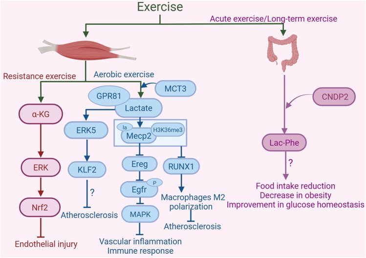
Atherosclerosis represents a complex interplay of inflammatory and metabolic processes, in which oxidative stress, endothelial inflammation, the phenotypic transition of smooth muscle cells (SMCs), and the conversion of macrophages into foam cells are involved. In contrast to pharmacological interventions, exercise emerges as a viable, cost-effective, and low-risk strategy to alleviate the progression of atherosclerosis. Exercise exerts beneficial effects on atherosclerosis through modulation of diverse pathways, including exerkines, browning of adipose tissue, the renin-angiotensin system (RAS), metabolites, gut microbiota, cell death pathways, microRNAs, nervous system, and immune function. The beneficial impacts of exercise on atherosclerosis and the mechanisms behind them will be examined here. Fully understanding the effects and mechanisms of exercise in reducing atherosclerosis might open doors to developing safe and effective interventions.

Keywords: atherosclerosis; browning of adipose tissue; cell death; exercise; exerkines; immune function; microRNAs.


Conflict of interest statement

The authors declare that the research was conducted in the absence of any commercial or financial relationships that could be construed as a potential conflict of interest.

Figures

FIGURE 1
FIGURE 1
Exercise-induced exerkines alleviate the development of atherosclerosis. Exercise increases the expression and sensitivity of fibroblast growth factor 21 (FGF21). FGF21 binding to FGFR1 triggers the PI3K/AKT pathway, suppresses IRE1α/JNK activation, and lowers levels of apoptosis proteins like CHOP and cleaved-caspase3, mitigating oxidative stress and apoptosis. In addition, exercise leads to an increase in the levels of glucagon-like peptide-1 (GLP-1) secreted by intestinal L cells. The activation of GLP-1R by GLP-1 can inhibit AKT to reduce mTOR activity, which helps to control the inflammatory response in human coronary artery SMCs. Exercise upregulates the expression of adiponectin, which increases NO content by promoting AMPKα1/AMPKα2 expression, consequently inhibiting endothelial activation. Furthermore, exercise also increases the production of irisin, which can inhibit the expression of PCSK9 via AMPK/SREBP2 signaling pathway, leading to a reduction in inflammation and plaque instability. Moreover, exercise can decrease the expression of TNFα to inhibit inflammation and the instability of atherosclerotic plaques. Additionally, exercise can suppress the expression of resistin to inhibit the p38MAPK/AP-1 signaling pathway, thereby alleviating inflammation and the plaque instability associated with atherosclerosis. Exercise can also inhibit the expression of leptin, suppressing JAK/STAT and MAPK signaling pathways, thereby alleviating inflammation and plaque instability. The graph was created with biorender.com (agreement number: IE28KV91XQ).
FIGURE 2
FIGURE 2
Exercise-regulated renin-angiotensin system (RAS) in amelioration of atherosclerosis. Under conditions of atherosclerosis, renin initiates the hydrolysis of angiotensinogen (ATG) to generate angiotensin I (Ang I), which is subsequently converted to angiotensin II (Ang II) by angiotensin-converting enzyme (ACE). This represents the activation of the classical arm of the renal RAS. Exercise inhibits the production of ACE and the binding of Ang II to the receptor AT1R, thereby reducing the activation of the JNK/ERK signaling pathway and suppressing early growth response factor 1 (Egr-1) expression, and consequently mitigating vas-oconstriction, inflammation, oxidative stress, and cell adhesion. Additionally, exercise can enhance the expression of AT2R in the heart. AT2R binds to Ang III, which is generated from Ang II by the action of aminopeptidase A (APA), thereby exerting diuretic, vasodilatory, and anti-inflammatory effects. Exercise also promotes the production of ACE2, which facilitates the conversion of Ang II into angiotensin-(1-7) [Ang 1-7], activating the non-classical pathway of the RAS. Ang 1-7 is a vasodilator peptide that exerts anti-inflammatory, antioxidative, and antiproliferative actions by binding to the receptor Mas, thereby alleviating the development of atherosclerosis. The graph was created with biorender.com (agreement number: HW28KV9DFS).
FIGURE 3
FIGURE 3
Exercise-induced metabolites in the protection against atherosclerosis. Resistance exercise enhances the synthesis and release of α-ketoglutarate (α-KG) in mouse muscle. α-KG activates nuclear factor erythroid 2-related factor 2 (Nrf2) by activating the ERK signaling pathway, inhibiting oxidative stress and mitochondrial dysfunction, thereby providing protective effects against endothelial damage. Aerobic exercise increases the production of lactate in skeletal muscle. Monocarboxylate transporter 3 (MCT3) is involved in the transport of lactate out of skeletal muscle. Lactate, by binding to the receptor GPR81, activates the ERK5 pathway to increase the expression of the arterial anti-atherosclerotic protective transcription factor Kruppel-like factor 2 (KLF2), thus playing a role in resisting atherosclerosis. Furthermore, lactate can activate lactylation of Mecp2 on lysine 271 in aortic ECs, subsequently inhibiting the expression of epidermal growth factor receptor (Egfr) ligand epiregulin (Ereg). This inhibition reduces the phosphorylation of Egfr, thereby blocking its downstream MAPK signaling pathway. These changes collectively alleviate vascular inflammation and immune responses. Additionally, exercise promotes the lactylation modification of Mecp2 at lysine 271, which facilitates the interaction of histone H3 trimethylation at Lys36 (H3K36me3) in macrophages of the aortic root plaque. This interaction leads to the demethylation of H3K36me3, suppression of the expression of RUNX family transcription factor 1 (RUNX1), and promotion of M2 macrophage polarization, thereby alleviating atherosclerosis. Acute exercise and long-term exercise training promote the production of Lac-Phe in macrophages and intestinal epithelial cells, with CNDP2 being a major Lac-Phe biosynthetic enzyme. The increase in Lac-Phe is associated with reduced food intake, decreased obesity, and improved glucose homeostasis, which may contribute to the alleviation of atherosclerosis. The graph was created with biorender.com (agreement number: KL28KV9IFU).
FIGURE 4
FIGURE 4
Exercise-mediated gut microbiota changes in the amelioration of atherosclerosis. Exercise can alter the composition of the gut microbiota, increasing the abundance of Bacteroidetes, Blautia, Rikenellaceae, and Dubosiella, while decreasing the abundance of Desulfovibrio, Tyzzerella, Lachnospiraceae-ge, and Gammaproteobacteria. By regulating the gut microbiota, exercise inhibits the binding of lipopolysaccharide (LPS) and Toll-like receptor 4 (TLR4), suppressing the MyD88 signaling pathway, which in turn suppresses inflammatory responses and the formation of atherosclerotic plaques. Additionally, exercise inhibits trimethylamine N-oxide (TMAO) expression and the NLR family pyrin domain-containing 3 (NLRP3) inflammasome to suppress inflammatory responses, thereby promoting the stability of atherosclerotic plaques. Furthermore, exercise can inhibit branched-chain amino acids (BCAAs) production and promote short-chain fatty acid (SCFA) to exert anti-atherosclerotic effects. The graph was created with biorender.com (agreement number: UF28KV9P6Z).
FIGURE 5
FIGURE 5
Exercise-regulated cell death in amelioration of atherosclerosis. Exercise may mitigate ferroptosis by activating AMPKα2, reducing oxidative stress, and inhibiting lipid peroxidation, ultimately alleviating atherosclerosis. Exercise can enhance the expression of glutaminase (GLS) to produce antioxidant glutathione (GSH), which can reduce Cu (II) to Cu (I). Subsequently, it forms inactive complexes by binding with Cu (I), thereby preventing copper ions from disrupting the normal structure and function of bovine serum albumin (BSA) within cells, thus inhibiting cuproptosis and potentially mitigating atherosclerosis. Moreover, exercise can reduce the expression of nuclear paraspeckle assembly transcript 1 (NEAT1) in the thoracic aorta of atherosclerotic mice. The downregulation of NEAT1 disrupts the transcriptional activation of the downstream gene NLRP3 by kruppel-like factor 4 (KLF4), leading to decreased expression of NLRP3. This reduction in NLRP3 expression results in a decrease in the expression of the NLRP3-mediated pyroptosis-associated gene GSDMD, which contributes to the inhibition of EC pyroptosis and thus alleviates atherosclerosis. In addition, exercise may reduce the activation of the JNK signaling pathway induced by endoplasmic reticulum (ER) stress, thereby inhibiting apoptosis and alleviating atherosclerosis. The graph was created with biorender.com (agreement number: KB28KV9VWL).
FIGURE 6
FIGURE 6
Exercise-regulated miRNAs, nervous system, immune cells, and m6A in the alleviation of atherosclerosis. Exercise alleviates atherosclerosis by modulating the expression of miRNAs, which target genes to promote translational repression or degradation, thereby inhibiting osteogenic programs and vascular calcification, VSMCs proliferation and migration, lipid accumulation and oxidative stress, as well as cell apoptosis and inflammation. Additionally, exercise mitigates atherosclerosis by promoting parasympathetic activity and inhibiting sympathetic activity, thereby suppressing the expression of norepinephrine (NE) and MAO-A, and reducing mitochondrial dysfunction, oxidative stress, and inflammation. Furthermore, exercise may maintain the homeostasis of the immune microenvironment by inhibiting the release of neutrophil extracellular traps (NETs) from neutrophils and the production of TNFα by B2 cells, promoting the generation of natural IgM antibodies from B1 cells and the secretion of IL-10 by Tregs, as well as suppressing the inhibitory effects of dendritic cells (DCs) on Tregs, thereby alleviating atherosclerosis. Exercise also diminishes METTL14 activity and expression in the thoracic aorta of atherosclerotic mice. This inhibition reduces METTL14-mediated m6A modification of NEAT1 mRNA, leading to lower NEAT1 expression, which ultimately suppresses endothelial cell pyroptosis and alleviates atherosclerosis. The graph was created with biorender.com (agreement number: QK28KVA0MB).

References

    1. Adkar S. S., Leeper N. J. (2024). Efferocytosis in atherosclerosis. Nat. Rev. Cardiol. 21, 762–779. 10.1038/s41569-024-01037-7 - DOI - PubMed
    1. Aguilar-Ballester M., Herrero-Cervera A., Vinue A., Martinez-Hervas S., Gonzalez-Navarro H. (2020). Impact of cholesterol metabolism in immune cell function and atherosclerosis. Nutrients 12 (7), 2021. 10.3390/nu12072021 - DOI - PMC - PubMed
    1. Ahmadi N., Eshaghian S., Huizenga R., Sosnin K., Ebrahimi R., Siegel R. (2011). Effects of intense exercise and moderate caloric restriction on cardiovascular risk factors and inflammation. Am. J. Med. 124 (10), 978–982. 10.1016/j.amjmed.2011.02.032 - DOI - PubMed
    1. Ai Y., Meng Y., Yan B., Zhou Q., Wang X. (2024). The biochemical pathways of apoptotic, necroptotic, pyroptotic, and ferroptotic cell death. Mol. Cell 84 (1), 170–179. 10.1016/j.molcel.2023.11.040 - DOI - PubMed
    1. Ajoolabady A., Pratico D., Lin L., Mantzoros C. S., Bahijri S., Tuomilehto J., et al. (2024). Inflammation in atherosclerosis: pathophysiology and mechanisms. Cell Death and Dis. 15 (11), 817. 10.1038/s41419-024-07166-8 - DOI - PMC - PubMed

LinkOut - more resources