Review
. 2025 Aug 11:15:1634909.
doi: 10.3389/fcimb.2025.1634909. eCollection 2025.

Mechanisms and therapeutic perspectives of mitochondrial dysfunction of macrophages in periodontitis

Affiliations
Review

Mechanisms and therapeutic perspectives of mitochondrial dysfunction of macrophages in periodontitis

Yibing Jia et al. Front Cell Infect Microbiol. .

Abstract

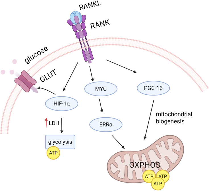
Periodontitis is a global inflammatory oral disease, and plaque-induced host excessive immune response is recognized as a major cause of its pathogenesis. In recent years, the relevance of mitochondrial dysfunction to periodontitis has been increasingly investigated, particularly with respect to macrophages, the key immune cells in the periodontal immune microenvironment. Mitochondrial dysfunction drives macrophage M1 polarization and osteoclast differentiation through mechanisms such as metabolic reprogramming, reactive oxygen species release, abnormal mitophagy, abnormal mitochondrial biogenesis and damaged mitochondrial dynamic. In addition, mitochondrial transfer in the periodontitis setting has been reported in several researches. In this review, we highlight the impact of mitochondrial dysfunction on macrophages in the periodontitis setting and summarize emerging therapeutic strategies for targeting mitochondria in periodontitis, including antioxidants, modulators of metabolic reprogramming, nanomaterials and photodynamic therapy.

Keywords: macrophage polarization; mitochondrial dysfunction; osteoclast differentiation; periodontitis mechanism; periodontitis treatment.


Conflict of interest statement

The authors declare that the research was conducted in the absence of any commercial or financial relationships that could be construed as a potential conflict of interest. The reviewer HH declared a shared affiliation with the authors to the handling editor at the time of review.

Figures

Figure 1
Figure 1
Comparison of cellular components between periodontitis and normal tissues. The left side indicates healthy periodontal tissue and the right side indicates periodontitis. It can be found that compared to healthy periodontal tissue, the proportion of M1 macrophages is up-regulated. The proportion of M2 macrophages decreases in periodontitis conditions. There is an imbalance between osteoblasts and osteoclasts of the alveolar bone, manifested by greater bone resorption than bone formation.
Figure 2
Figure 2
Regulation of mitochondrial core dynamic process. (A) Schematic diagram of mitochondrial fission, Drp1 is the main fission regulator. (B) Schematic diagram of mitophagy, PINK1 accumulates in the outer mitochondrial membrane (OMM) and recruits Parkin to the mitochondrial surface, which in turn triggers mitophagy. (C) Schematic diagram of mitochondrial genesis, PGC-1α/β acts as a general regulator of mitochondrial biogenesis by activating the transcription of downstream genes. (D) Schematic diagram of mitochondrial fusion, Mfn1 and Mfn2-mediated fusion of the OMM, and OPA1 control of the inner mitochondrial membrane fusion.
Figure 3
Figure 3
Mitochondrial metabolism in M2 and M1 macrophages: TCA cycle and OXPHOS. The left part represents the TCA cycle in the mitochondria of M2 macrophages with the mtETC-OXPHOS system. As the figure shows, the TCA cycle is normal in M2 macrophages. The mtETC-OXPHOS system exists in the inner membrane of the mitochondria, and the OXPHOS system consists of four complexes, ATP synthase and two mobile electron carriers (CoQ and Cytc). Among them, complexes I, III and IV have a proton pump function and are responsible for pumping protons from the mitochondrial matrix to the membrane interstitial space to form a proton gradient. While complex II is not involved in proton pumping and only transfers electrons to CoQ. ATP synthase uses the proton motive force potential generated by the ETC to pump protons from the membrane gap back to the mitochondrial matrix, while phosphorylating ADP to ATP, which provides energy to the cell. In contrast, right-sided M1 macrophages have impaired TCA cycling, which in turn affects mtETC efficiency and reduces oxidative phosphorylation. In addition, M1 macrophages showed reverse electron transfer (RET), resulting in increased electron leakage and increased mtROS production.
Figure 4
Figure 4
RANKL signaling regulates osteoclast differentiation. RANKL signaling promotes OXPHOS and ATP production by inducing PGC1β, which is involved in mitochondrial biogenesis, and the MYC-ERRα axis. RANKL also increases GLUT number and glycolysis by up-regulating HIF-1α during the later stages of osteoclast differentiation, thereby increasing glucose uptake and utilization.
Figure 5
Figure 5
Downstream pathways of mtROS release. Activation of MAPK and NF-κB pathways (e.g. activation of p38 into the nucleus, up-regulation of IκBα transcription, etc.), which in turn elicits an inflammatory response. Stabilization of HIF-1α, which facilitates aerobic glycolysis and IL-1β induction in LPS-activated macrophages. Activation of the NLRP3 inflammatory vesicle, activation of Caspase-1, cleavage of pro-IL-1β and pro-IL-18 to transform them to active forms.

Similar articles

References

    1. Abraham D., Singh A., Goyal A. (2023). Salivary levels of NLRP3 protein are significantly raised in chronic periodontitis: A systematic review and meta-analysis of clinical studies. J Indian Soc Periodontol. 27, 552–558. doi: 10.4103/jisp.jisp_185_23, PMID: - DOI - PMC - PubMed
    1. Adam-Vizi V., Chinopoulos C. (2006). Bioenergetics and the formation of mitochondrial reactive oxygen species. Trends Pharmacol. Sci. 27, 639–645. doi: 10.1016/j.tips.2006.10.005, PMID: - DOI - PubMed
    1. Adebayo M., Singh S., Singh A. P., Dasgupta S. (2021). Mitochondrial fusion and fission: The fine-tune balance for cellular homeostasis. FASEB J. 35, e21620. doi: 10.1096/fj.202100067R, PMID: - DOI - PMC - PubMed
    1. Adhikari N., Prasad Aryal Y., Jung J. K., Ha J. H., Choi S. Y., Kim J. Y., et al. (2021). Resveratrol enhances bone formation by modulating inflammation in the mouse periodontitis model. J. Periodontal Res. 56, 735–745. doi: 10.1111/jre.12870, PMID: - DOI - PubMed
    1. Ahn H., Lee K., Kim J. M., Kwon S. H., Lee S. H., Lee S. Y., et al. (2016). Accelerated lactate dehydrogenase activity potentiates osteoclastogenesis via NFATc1 signaling. PLoS One 11, e0153886. doi: 10.1371/journal.pone.0153886, PMID: - DOI - PMC - PubMed

LinkOut - more resources