

The Great Mimicker: Extragonadal Endometriosis Presenting as an Appendiceal Mass and Acute Appendicitis
- PMID: 40861586
- PMCID: PMC12375226
- DOI: 10.7759/cureus.88718
The Great Mimicker: Extragonadal Endometriosis Presenting as an Appendiceal Mass and Acute Appendicitis
Abstract
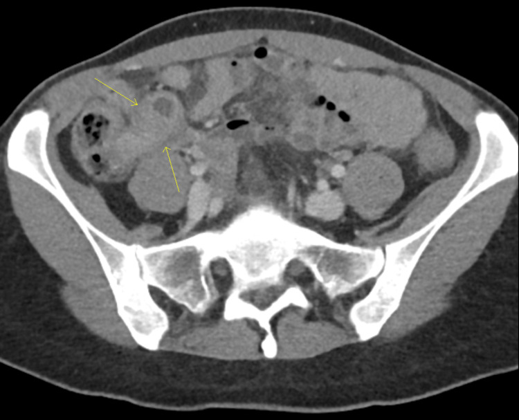
A 45-year-old female presented to the Emergency Department with acute right lower quadrant (RLQ) pain. Her past medical history was significant for heavy menstrual bleeding and moderate dysmenorrhea, though a diagnosis of endometriosis had never been established previously. Initial work-up revealed anemia and leucocytosis. A CT of the abdomen and pelvis showed an enlarged structure in RLQ along the medial border of the cecum, suspicious for acute appendicitis with a retained rupture. CA19-9, CA-125, and CEA were sent, of which only CA-125 was elevated at 92.7 U/mL (reference range: 0-35 U/mL). She was admitted to the hospital for complicated appendicitis and was started on IV piperacillin-tazobactam. A repeat CT scan to monitor response to antibiotics showed acute appendicitis along with phlegmon with a small amount of free fluid, and an enlarged heterogeneous uterus with thickened endometrium, uterine fibroids, and a left ovarian cyst. A CT-guided drainage by Interventional Radiology (IR) was deferred due to concerns for a suspected malignant appendiceal mass, and she was treated with 14 days of antibiotics in the setting of complicated appendicitis. An appendiceal biopsy via colonoscopy after discharge revealed a serrated adenoma. The patient underwent a laparoscopic appendectomy and a right hemicolectomy due to concern for appendiceal malignancy. The final histopathology showed a 2.8 cm endometriotic mass involving the wall of the appendix and surrounding tissue, one diverticulum of the colon, and was negative for dysplasia and malignancy. She had a further gynecologic workup with an in-office hysteroscopy, endometrial biopsy, and a pelvic MRI. The pathology showed no evidence of atypical hyperplasia or malignancy. The MRI revealed numerous uterine leiomyomas, adenomyosis, and a left adnexal endometrioma measuring 3.6 x 3.2 cm. She underwent a subsequent robotic-assisted total hysterectomy, bilateral salpingectomy, left oophorectomy, right ovarian cystectomy, and extensive adhesiolysis/enterolysis with intraoperative colorectal surgery consultation. Her post-operative course was uncomplicated. This report highlights the importance of considering extragonadal endometriosis in women of reproductive age as a differential diagnosis for RLQ pain and mass. It also underscores the importance of investigating the preoperative findings of appendiceal masses in women of childbearing age with appendiceal endometriosis presenting as an acute abdomen.
Keywords: acute abdomen; appendectomy; appendiceal endometriosis; appendiceal mass; appendicitis; endometriosis; extragonadal endometriosis; right lower quadrant abdominal pain.
Copyright © 2025, Ha et al.
Conflict of interest statement
Human subjects: Informed consent for treatment and open access publication was obtained or waived by all participants in this study. Conflicts of interest: In compliance with the ICMJE uniform disclosure form, all authors declare the following: Payment/services info: All authors have declared that no financial support was received from any organization for the submitted work. Financial relationships: All authors have declared that they have no financial relationships at present or within the previous three years with any organizations that might have an interest in the submitted work. Other relationships: All authors have declared that there are no other relationships or activities that could appear to have influenced the submitted work.
Figures


References
Publication types
LinkOut - more resources
Full Text Sources
Research Materials
Miscellaneous