. 2025 Aug 20;15(8):549.
doi: 10.3390/bios15080549.

Preliminary Study Using Wearable Near-Infrared Spectroscopy for Continuous Monitoring of Hemodynamics Through the Carotid Artery

Affiliations

Preliminary Study Using Wearable Near-Infrared Spectroscopy for Continuous Monitoring of Hemodynamics Through the Carotid Artery

Nisha Maheshwari et al. Biosensors (Basel). .

Abstract

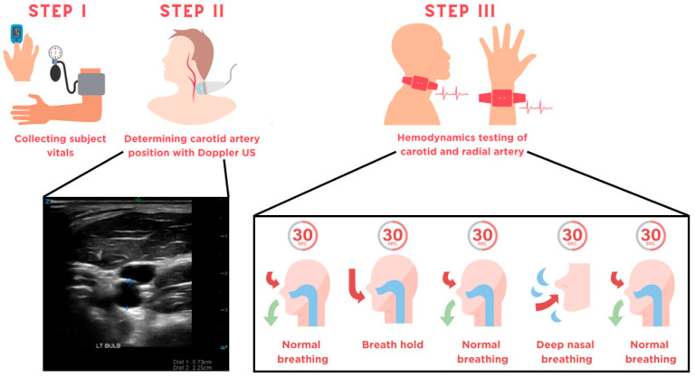
Non-invasive, continuous monitoring of carotid artery hemodynamics may provide valuable insights on cerebral blood perfusion (CBP). Near-infrared spectroscopy (NIRS) is a non-invasive modality that may be a good candidate for real-time carotid artery monitoring. We designed a wearable NIRS system to monitor the left and right radial and carotid arteries in 20 healthy subjects. The changes in total hemoglobin concentration (HbT) and tissue oxygen saturation (StO2) in all 80 arteries were continuously monitored in response to changes in oxygen supply. Wilcoxon non-parametric equivalence testing was used to compare changes in the radial (reference) and carotid arteries. The system-derived HbT and StO2 trends matched the expected physiological responses over time in the radial and carotid arteries. The mean peak-to-peak amplitude [uM] of HbT during sustained deep breathing was practically equivalent between the left radial (0.9 ± 0.8) and left carotid (1.6 ± 1.1) arteries (p = 0.01). The mean peak-to-peak amplitude [%] of StO2 was practically equivalent between the left radial (0.3 ± 0.2) and left carotid (0.3 ± 0.2) arteries (p < 0.001) and the right radial (0.4 ± 0.5) and right carotid (0.5 ± 0.4) arteries (p = 0.001). These findings indicate that NIRS may be a good option for monitoring the carotid arteries to track changes in CBP.

Keywords: Biophotonics; carotid artery; hemodynamics; near-infrared spectroscopy; tissue oxygen saturation.


Conflict of interest statement

All authors are inventors on PCT patent application no. PCT/US2024/038690. NM and LS own shares of CaroRhythm, Inc, and NM is the acting CEO of CaroRhythm, Inc.

Figures

Figure 1
Figure 1
(a) Patch that sits over the left and right radial arteries. (b) Patch that sits over the left and right carotid arteries. Both patches are 60 mm × 25 mm in size and have the same source-detector distances of 25 mm and 32 mm.
Figure 2
Figure 2
Infographic detailing the data acquisition protocol. In Step I, the heart rate, blood pressure, and oxygen saturation are taken in the supine position. In Step II, transverse images of the carotid bulb, internal carotid artery, and external carotid artery on the left and right sides of the subject are taken. Finally, in Step III the subject was instructed to perform a series of breathing exercises, during which their left and right radial and carotid arteries were monitored with the NIRS system.
Figure 3
Figure 3
(a) The change in HbT [uM] from baseline over time for a representative trial. The breath hold (BH) and deep breathing (DB) segments of the trial are indicated. The red star marks the maximum change in HbT [uM] during the BH. The red dots mark a characteristic peak-to-peak amplitude [uM] of one oscillation. (b) The change in StO2 [%] from baseline over time for a representative trial. The BH and DB segments are indicated. The red dots mark a characteristic time between oscillations [sec]. For clarity, the left radial artery was used as an example to highlight parameters of interest, and all parameters were calculated for both features (HbT and StO2) at all locations.
Figure 4
Figure 4
The maximum percentage change in (a) HbT [%] and (b) StO2 [%], the mean peak-to-peak amplitude of (c) HbT [uM] and (d) StO2 [%], and the mean oscillation time for (e) HbT [sec] and (f) StO2 [sec]. The following arteries are compared for equivalency: (1) the left radial and left carotid; (2) the right radial and right carotid; (3) the left and right radials; and (4) the left and right carotids. Arteries were compared using the Wilcox-TOST equivalence test. n.s. = no significant equivalence; *= p ≤ 0.05; **= p < 0.01; ***= p < 0.001.

References

    1. Prabhakar N.R. Oxygen Sensing by the Carotid Body Chemoreceptors. J. Appl. Physiol. 2000;88:2287–2295. doi: 10.1152/jappl.2000.88.6.2287. - DOI - PubMed
    1. Del Rio R., Moya E.A., Iturriaga R. Carotid Body Potentiation during Chronic Intermittent Hypoxia: Implication for Hypertension. Front. Physiol. 2014;5:434. doi: 10.3389/fphys.2014.00434. - DOI - PMC - PubMed
    1. Sweid A., Hammoud B., Ramesh S., Wong D., Alexander T.D., Weinberg J.H., Deprince M., Dougherty J., Maamari D.J.-M., Tjoumakaris S., et al. Acute Ischaemic Stroke Interventions: Large Vessel Occlusion and Beyond. Stroke Vasc. Neurol. 2019;5:80–85. doi: 10.1136/svn-2019-000262. - DOI - PMC - PubMed
    1. Phan T.G., Beare R.J., Jolley D., Das G., Ren M., Wong K., Chong W., Sinnott M.D., Hilton J.E., Srikanth V. Carotid Artery Anatomy and Geometry as Risk Factors for Carotid Atherosclerotic Disease. Stroke. 2012;43:1596–1601. doi: 10.1161/STROKEAHA.111.645499. - DOI - PubMed
    1. Qaja E., Tadi P., Theetha Kariyanna P. StatPearls. StatPearls Publishing; Treasure Island, FL, USA: 2025. Symptomatic Carotid Artery Stenosis. - PubMed

LinkOut - more resources