. 2025 Aug 29;11(35):eady8168.
doi: 10.1126/sciadv.ady8168. Epub 2025 Aug 27.

Human endogenous retrovirus K (HERV-K) envelope structures in pre- and postfusion by cryo-EM

Affiliations

Human endogenous retrovirus K (HERV-K) envelope structures in pre- and postfusion by cryo-EM

Jeremy Shek et al. Sci Adv. .

Abstract

Human endogenous retroviruses (HERVs) are remnants of ancient infections that comprise ~8% of the human genome. The HERV-K envelope glycoprotein (Env) is aberrantly expressed in cancers, autoimmune disorders, and neurodegenerative diseases, and is targeted by patients' own antibodies. However, a lack of structural information has limited molecular and immunological studies of the roles of HERVs in disease. Here, we present cryo-electron microscopy structures of stabilized HERV-K Env in the prefusion conformation, revealing a distinct fold and architecture compared to HIV and simian immunodeficiency virus. We also generated and characterized a panel of monoclonal antibodies with subunit and conformational specificity, serving as valuable research tools. These antibodies enabled structure determination of the postfusion conformation of HERV-K Env, including its unique "tether" helix, and antibody-bound prefusion Env. Together, these results provide a structural framework that opens the door to mechanistic studies of HERV-K Env and tools for its evaluation as a potential therapeutic target.


Figures
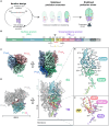
Fig. 1.
Fig. 1.. Stabilized prefusion HERV-K Env enables high-resolution structure determination by cryo-EM trimer.
(A) Protein engineering workflow for stabilization of the prefusion HERV-K EnvECTO. (B) Primary structure annotation of EnvECTO construct. Domains are color-coded. Abbreviations: FP, fusion peptide; HR1, heptad repeat 1; HR2, heptad repeat 2; GS-Link, 12-amino acid GGS linker; T4-Fib, T4 fibritin foldon trimerization domain; 2xST, Twin Strep-Tag II. Regions boxed in black are sequences of EnvECTO. Black scissors indicate the furin cleavage site that cleaves Env into its SU and TM subunits. Engineered disulfide bond mutations and furin-cleavage motif modifications are annotated. (C) 2.2-Å resolution cryo-EM map of the HERV-K EnvECTO trimer (colored using green, red, and blue shades) (D) Cartoon representation of the HERV-K EnvECTO trimer. Protomer A is colored in a rainbow as described in (B). The remaining two protomers are colored in white. (E) Isolated cartoon representation of the SU subunit from an individual protomer. (F) Cartoon representation of the TMECTO. Both (E) and (F) are colored using the same color scheme as (B) and (D). N- and C-terminal Cα atoms for each subunit are represented as spheres.
Fig. 2.
Fig. 2.. Protomer and trimer inferfaces of prefusion Env.
(A) The prefusion EnvECTO trimer displayed using a molecular surface representation. SU + TM protomer interactions are shown in (B to E) and trimeric interactions are shown in (F) and (G). (B) The clasp formed by the TM around the base domain of the SU viewed from the front. Residues involved in clasp locking are shown as sticks. (C) The C-shaped clasp formed by TM around SU viewed from bottom-up. [(B) and (C)] SU is shown as a light green surface and TM is a dark green cartoon. (D) β Sheet formation between the β1 strand from SU (light green) and the β27 strand from TM (dark green). (E) The engineered disulfide bond between the SU and TM to stabilize EnvECTO. (F) Side view of the central coiled coil. Hydrophobic residues facing the interface from each helix are shown as sticks. (G) The interactions between adjacent prefusion TM subunits distal from the central coils. The fusion peptide of TMA is colored in yellow. [(B), (D), and (G)]. Hydrogen bond interactions are shown by dashed black lines, and salt bridges are shown in dashed yellow lines.
Fig. 3.
Fig. 3.. Prefusion HERV-K Env has a unique fold compared to previously solved prefusion retroviral structures.
(A) Models of HERV-K Env, HIV Env (closed (PDB:5CEZ), occluded (PDB:7TFO), open (PDB:5VN3), and SIV Env (PDB:7T4G) ectodomain trimers. Approximate heights and widths of each complex were measured using ChimeraX (56). (B) Comparison of receptor-binding subunits (SU: HERV-K, Syncytin-2; gp120: HIV, SIV). HERV-K Env SU is colored by subdomain, while the other receptor binding subunits are colored in light gray. Shared structural features to HERV-K SU are colored and labeled with their respective colors. (C) Comparison of fusion protein subunits (TM: HERV-K; gp41: HIV, SIV) in prefusion conformation. HERV-K Env TM is colored by subdomain. Similar structural features presented on the HIV and SIV gp41 subunits are colored similarly to TM. The C-shaped clasp that encloses the SU/gp120 base domain is boxed in black on each TM/gp41. The HR1 central coil in the occluded and closed conformations of gp41 are extended at the N-terminal ends of the helix, highlighted by the dashed box. Calculated all-atom RMSD values against the HERV-K Env complex or respective subunit are displayed under each structure.
Fig. 4.
Fig. 4.. mAbs with subunit and conformational specificity enable detection of Env in patient-derived samples.
(A) Binding of mAbs to unstabilized HERV-K Phoenix EnvUNS (EnvUNS; blue), denatured EnvUNS (denatured EnvUNS; green), the MBP-TMECTO construct (postfusion TMECTO; pink), and denatured MBP-TMECTO (denatured TMECTO; orange) determined by ELISA. Each mAb was run in duplicate (n = 2) and each data point plotted as a circle. (B) Western blots using anti-Env mAbs as the primary antibody. All blots used purified, unstabilized EnvUNS, which contains both SU (~51 kDa) and TMECTO (~30 kDa) bands. (C) Table of anti-Env mAbs found in this study. Antibodies were screened for SU or TM binding using ELISA shown in fig. S1A. Epitope type was determined using ELISAs in combination with Western blots. * = presumed linear based on Western blotting results. (D) Immunofluorescence staining of isolated neutrophils from either SLE, RA, or healthy donors using mAbs from this study and the commercially available TM-binding HERM-1811-5 antibody. Healthy neutrophils show no staining by any mAb.
Fig. 5.
Fig. 5.. Cryo-EM structure of the Kenv-4 bound postfusion HERV-K TMECTO complex reveals the 6HB architecture.
(A) Schematic representation of the postfusion HERV-K TMECTO construct. Abbreviations: GS-link, 17-amino acid linker. (B) 2.8-Å resolution cryo-EM map of postfusion TMECTO trimer (colored using green, blue, and red shades) bound to three copies of the Kenv-4 Fab. A low-resolution map is overlaid and represented as a silhouette. (C) A ribbon representation of the postfusion TMECTO. 6HB and central coiled-coil structural features are shown using dotted triangles. (D) A protomer of postfusion TMECTO with domains colored as described in (A).

References

    1. Paces J., Pavlícek A., Paces V., HERVd: Database of human endogenous retroviruses. Nucleic Acids Res. 30, 205–206 (2002). - PMC - PubMed
    1. Xue B., Sechi L. A., Kelvin D. J., Human endogenous retrovirus K (HML-2) in health and disease. Front. Microbiol. 11, 1690 (2020). - PMC - PubMed
    1. Nelson P. N., Carnegie P. R., Martin J., Davari Ejtehadi H., Hooley P., Roden D., Rowland-Jones S., Warren P., Astley J., Murray P. G., Demystified. Human endogenous retroviruses. Mol. Pathol. 56, 11–18 (2003). - PMC - PubMed
    1. Flockerzi A., Ruggieri A., Frank O., Sauter M., Maldener E., Kopper B., Wullich B., Seifarth W., Müller-Lantzsch N., Leib-Mösch C., Meese E., Mayer J., Expression patterns of transcribed human endogenous retrovirus HERV-K(HML-2) loci in human tissues and the need for a HERV transcriptome project. BMC Genomics 9, 354 (2008). - PMC - PubMed
    1. Yang C., Guo X., Li J., Han J., Jia L., Wen H.-L., Sun C., Wang X., Zhang B., Li J., Chi Y., An T., Wang Y., Wang Z., Li H., Li L., Significant upregulation of HERV-K (HML-2) transcription levels in human lung cancer and cancer cells. Front. Microbiol. 13, 850444 (2022). - PMC - PubMed

MeSH terms

LinkOut - more resources