. 2025 Aug 11;47(8):641.
doi: 10.3390/cimb47080641.

Molecular Insights into Tumor Immunogenicity

Affiliations

Molecular Insights into Tumor Immunogenicity

Irini Doytchinova et al. Curr Issues Mol Biol. .

Abstract

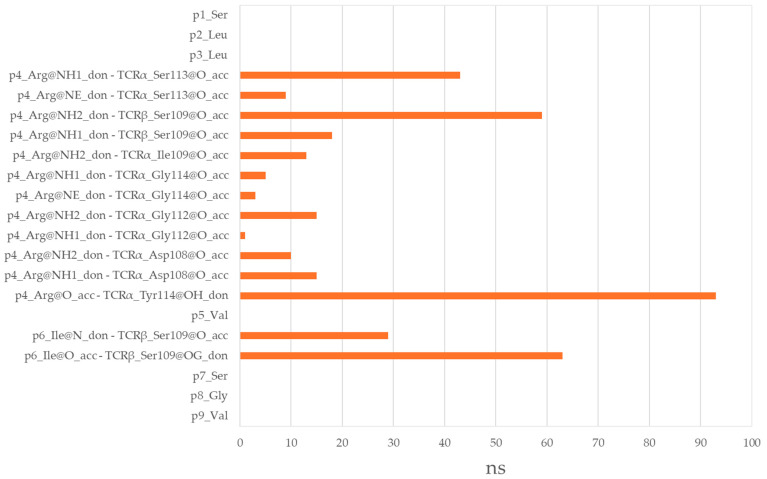
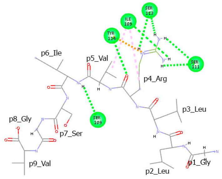
Tumor immunogenicity depends on the ability of peptides to form stable and specific interactions with both HLA molecules and T-cell receptors (TCRs). While HLA binding is essential, not all HLA-binding peptides elicit T-cell responses. This study investigates the molecular features distinguishing immunogenic T-cell epitopes from non-immunogenic HLA binders. Two datasets of nonamer peptides-38 T-cell epitopes and 144 non-epitopes-were compiled and analyzed using sequence logo models and molecular dynamics (MD) simulations of TCR-peptide-HLA complexes. A comparative logo analysis revealed strong amino acid preferences at central positions (p4-p8) in T-cell epitopes and absences in non-epitopes. A representative epitope-non-epitope pair was selected for structural modeling and 100 ns MD simulations. The T-cell epitope formed a more stable complex with the TCR and exhibited greater flexibility, supporting an induced-fit recognition mechanism. It also established a broader and longer-lasting network of hydrogen bonds and π interactions across the residues at positions p4-p8. In contrast, the non-epitope engaged TCR at only two positions. These findings highlight the critical role of the peptide's central region in TCR engagement and provide structural insights useful for neoantigen prediction, vaccine design, and TCR-based immunotherapies.

Keywords: HLA class I binders; T-cell epitopes; TCR-based immunotherapies; immunogenicity; molecular dynamics simulations; neoantigen prediction; vaccine design.


Conflict of interest statement

The authors declare no conflicts of interest.

Figures

Figure 1
Figure 1
Sequence logo for T-cell epitopes.
Figure 2
Figure 2
X-ray crystal structure of the HLA–peptide–TCR complex (PDB code: 6RPA). (Left): View across the peptide binding groove; (right): view along the peptide binding groove. The HLA molecule is shown in orange, β-microglobulin in gold, TCR chain D and E in yellow and light green, respectively, and the peptide in cyan.
Figure 3
Figure 3
Sequence logo for non-T-cell epitopes.
Figure 4
Figure 4
Average backbone RMSDs over 100 frames (100 ns) of the HLA–peptide–TCR complexes containing T-cell epitope SLLMWITQV (ink blue) and non-T-cell epitope GLLRVISGV (orange).
Figure 5
Figure 5
(a) Average RMSFs by residue of the HLA–peptide–TCR complexes containing T-cell epitope SLLMWITQV (blue shades) and non-T-cell epitope GLLRVISGV (brown shades). Residues 1–276 correspond to HLA protein, residues 277–375 to β-microglobulin, residues 376–384 to peptide (epitope given in green, non-epitope in red), residues 385–550 to TCRα, residues 551–785 to TCRβ. (b) Average RMSFs by residue of the bound peptide only.
Figure 6
Figure 6
Hydrogen bonds between the T-cell epitope SLLMWITQV and TCR α and β chains.
Figure 7
Figure 7
Intermolecular interactions between the T-cell epitope SLLMWITQV and the TCR α and β chains observed in the final conformation of the complex at the 100th ns. Hydrogen bonds are represented by green dashed lines, while π–alkyl and π–cation interactions are indicated by pink dots.
Figure 8
Figure 8
Hydrogen bonds between the non-T-cell epitope GLLRVISGV and TCR α and β chains.
Figure 9
Figure 9
Intermolecular interactions between the non-T-cell epitope GLLRVISGV and the TCR α and β chains observed in the final conformation of the complex at the 100th ns. Hydrogen bonds are represented by green dashed lines, while alkyl and π–cation interactions are indicated by pink and orange dots, respectively.

References

    1. Davis M.M., Bjorkman P.J. T-cell antigen receptor genes and T-cell recognition. Nature. 1988;334:395–402. doi: 10.1038/334395a0. - DOI - PubMed
    1. Rudolph M.G., Stanfield R.L., Wilson I.A. How TCRs bind MHCs, peptides, and coreceptors. Annu. Rev. Immunol. 2006;24:419–466. doi: 10.1146/annurev.immunol.23.021704.115658. - DOI - PubMed
    1. Peters B., Sette A. Generating quantitative models describing the sequence specificity of biological processes with the stabilized matrix method. BMC Bioinform. 2005;6:132. doi: 10.1186/1471-2105-6-132. - DOI - PMC - PubMed
    1. Paul S., Weiskopf D., Angelo M.A., Sidney J., Peters B., Sette A. HLA class I alleles are associated with peptide-binding repertoires of different size, affinity, and immunogenicity. J. Immunol. 2013;191:5831–5839. doi: 10.4049/jimmunol.1302101. - DOI - PMC - PubMed
    1. Rappuoli R., Bottomley M.J., D’Oro U., Finco O., De Gregorio E. Reverse vaccinology 2.0: Human immunology instructs vaccine antigen design. J. Exp. Med. 2016;213:469–481. doi: 10.1084/jem.20151960. - DOI - PMC - PubMed

LinkOut - more resources