. 2025 Aug 12;47(8):646.
doi: 10.3390/cimb47080646.

Linolenic Acid Inhibits Cancer Stemness and Induces Apoptosis by Regulating Nrf2 Expression in Gastric Cancer Cells

Affiliations

Linolenic Acid Inhibits Cancer Stemness and Induces Apoptosis by Regulating Nrf2 Expression in Gastric Cancer Cells

Jen-Lung Chen et al. Curr Issues Mol Biol. .

Abstract

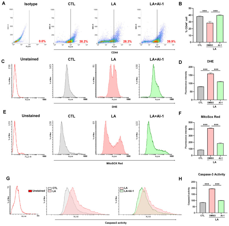
Although chemotherapy is the preferred treatment for gastric cancer, the therapeutic drugs currently available have limited efficacy and severe side effects. Cancer stem cells within tumor masses have the distinctive properties of self-renewal, maintenance, and resistance to chemotherapy. Hence, agents capable of targeting stemness in gastric tumors with minimal side effects are urgently required. Enzymes that generate reactive oxygen species contribute to the high oxidation levels observed in tumors. Additionally, nuclear factor erythroid 2-related factor 2 (Nrf2), an antioxidant transcription factor, regulates cancer stemness. Increasing evidence highlights the potential of nutritional supplementation to treat cancer stemness. ω-3 polyunsaturated fatty acids support human health and offer benefits for cancer treatment. Linolenic acid (LA), an ω-3 polyunsaturated fatty acid, inhibits the expression of proteins associated with stemness and promotes apoptosis in gastric cancer cells. Our findings indicated that LA treatment substantially inhibited key characteristics of gastric cancer stemness and induced oxidative stress and caspase-3-mediated apoptosis by downregulating Nrf2-mediated expression. These results suggest that LA is a promising nutritional supplement for targeting cancer stemness in the treatment of gastric cancer.

Keywords: Nrf2; cancer stemness; gastric cancer; linolenic acid; ω-3 polyunsaturated fatty acids.


Conflict of interest statement

The authors have no conflicts of interest to disclose.

Figures

Figure 1
Figure 1
(A) MKN45 cells were treated with linoleic acid (LA; 10, 25, or 50 μM) for 72 h. CD44+ cells were analyzed through flow cytometry. MKN45 cells were also stained with an isotype control antibody to serve as a background reference. (B) Quantification of CD44+ cells after LA (10, 25, or 50 μM) treatment. (C) Structural formula of LA. Data are presented as the mean ± standard error of the mean; n ≥ 3 independent experiments; two-tailed Student’s t test. *** p < 0.005.
Figure 2
Figure 2
(A) Nrf2 expression and (B) quantification of Nrf2 expression in nuclear fractions isolated from MKN45 cells, analyzed through Western blotting, after their treatment with LA (10, 25, or 50 μM) for 72 h. Lamin B1 served as the loading control. (C) SOX2 expression and (D) quantification of SOX2 expression in the nuclear fractions of MKN45 cells, analyzed through Western blotting, after their treatment with LA (10, 25, or 50 μM) for 72 h. Lamin B1 served as the loading control. (E) HO-1 expression and (F) quantification of HO-1 expression in MKN45 cells after their treatment with LA (10, 25, or 50 μM) for 72 h. (G,H) Mitochondrial oxidative stress in MKN45 cells, analyzed through mitoSOX red staining and flow cytometry, after their treatment with vehicle (CTL: black line) or LA (10, 25, or 50 μM; 10 μM: red line; 25 μM: blue line; 50 μM: green line)) for 72 h. The unstained cells (Unstained) serve as a background reference. The cells that were stained with mitoSOX red and treated with the vehicle act as the control group (CTL). (I,J) Reactive oxygen species generation in MKN45 cells, analyzed through dihydroethidium staining and flow cytometry, after their treatment with vehicle (CTL: black line) or LA (10, 25, or 50 μM; 10 μM: red line; 25 μM: blue line; 50 μM: green line) for 72 h. The unstained cells (Unstained) serve as a background reference. The cells that were stained with DHE and treated with the vehicle act as the control group (CTL). (K,L) Caspase-3 activity in MKN45 cells, analyzed through flow cytometry, after their treatment with LA (10, 25, or 50 μM) for 72 h. Data are presented as the mean ± standard error of the mean; n ≥ 3 independent experiments; two-tailed Student’s t test. * p < 0.05, ** p < 0.01, *** p < 0.005.
Figure 3
Figure 3
(A,B) CD44+ cell population in MKN45 cells, analyzed through flow cytometry, after their treatment with brusatol (40 nM) for 72 h. MKN45 cells were also stained with an isotype control antibody to serve as a background reference. (C) CD44, Nrf2, SOX2, HO-1, and β-actin expression and (D) quantification of the expression of these proteins in MKN45 cells after their treatment with brusatol (40 nM) for 72 h. (E,F) ROS generation in MKN45 cells, analyzed through dihydroethidium staining and flow cytometry, after their treatment with brusatol (40 nM) for 72 h. (G,H) Mitochondrial oxidative stress in MKN45 cells, analyzed through mitoSOX red staining and flow cytometry, after their treatment with brusatol (40 nM) for 72 h. (I,J) Caspase-3 activity in MKN45 cells, analyzed through flow cytometry, after their treatment with brusatol (40 nM) for 72 h. Data are presented as the mean ± standard error of the mean; n ≥ 3 independent experiments; two-tailed Student’s t test ** p < 0.01, *** p < 0.005.
Figure 4
Figure 4
(A,B) CD44+ cell population, analyzed through flow cytometry, in MKN45 cells treated with an LA (25 μM) vehicle, or an LA (25 μM) vehicle + AI-1 (10 μM) for 72 h. MKN45 cells were also stained with an isotype control antibody to serve as a background reference. (C,D) ROS generation, (E,F) mitochondrial oxidative stress, and (G,H) caspase-3 activation, observed through flow cytometry analysis, in MKN45 cells treated with an LA (25 μM) vehicle or an LA (25 μM) vehicle + Nrf2 activator AI-1 (10 μM) for 72 h. Data are presented as the mean ± standard error of the mean; n ≥ 3 independent experiments; two-tailed Student’s t test. *** p <0.005.
Figure 5
Figure 5
(A) Expression of phosphorylated-Akt and total Akt in MKN45 cells, analyzed through Western blotting, after their treatment with LA (25 μM) for 72 h. (B) Quantification of the phosphorylated-Akt/Akt ratio after cells’ treatment with LA (25 μM). (C,D) Nrf2 and Lamin B1 expression in nuclear fractions isolated from MKN45 cells and analyzed through Western blotting after the cells’ treatment with Akt inhibitor LY294002 (10 μM) for 1 h. (E) Nrf2 expression and (F) quantification of Nrf2 expression in MKN45 cells after their treatment with LY294002 (10 μM) for 72 h. Data are presented as the mean ± standard error of the mean; n ≥ 3 independent experiments; two-tailed Student’s t test. ** p < 0.01.

References

    1. Ilic M., Ilic I. Epidemiology of stomach cancer. World J. Gastroenterol. 2022;28:1187–1203. doi: 10.3748/wjg.v28.i12.1187. - DOI - PMC - PubMed
    1. Yang W.J., Zhao H.P., Yu Y., Wang J.H., Guo L., Liu J.Y., Pu J., Lv J. Updates on global epidemiology, risk and prognostic factors of gastric cancer. World J. Gastroenterol. 2023;29:2452–2468. doi: 10.3748/wjg.v29.i16.2452. - DOI - PMC - PubMed
    1. Marin J.J.G., Perez-Silva L., Macias R.I.R., Asensio M., Peleteiro-Vigil A., Sanchez-Martin A., Cives-Losada C., Sanchon-Sanchez P., Sanchez De Blas B., Herraez E., et al. Molecular Bases of Mechanisms Accounting for Drug Resistance in Gastric Adenocarcinoma. Cancers. 2020;12:2116. doi: 10.3390/cancers12082116. - DOI - PMC - PubMed
    1. Li Y.T., Tan X.Y., Ma L.X., Li H.H., Zhang S.H., Zeng C.M., Huang L.N., Xiong J.X., Fu L. Targeting LGSN restores sensitivity to chem-otherapy in gastric cancer stem cells by triggering pyroptosis. Cell Death Dis. 2023;14:545. doi: 10.1038/s41419-023-06081-8. - DOI - PMC - PubMed
    1. Lee C.H., Tsai H.Y., Chen C.L., Chen J.L., Lu C.C., Fang Y.P., Wu D.C., Huang Y.B., Lin M.W. Isoliquiritigenin Inhibits Gastric Cancer Stemness, Modulates Tumor Microenvironment, and Suppresses Tumor Growth through Glucose-Regulated Protein 78 Down-regulation. Biomedicines. 2022;10:1350. - PMC - PubMed

LinkOut - more resources