. 2025 Aug;10(8):101281.
doi: 10.1016/j.jacbts.2025.03.013.

Evaluation of Tideglusib as a Disease Modifying Therapy in Murine Models of Arrhythmogenic Cardiomyopathy

Affiliations

Evaluation of Tideglusib as a Disease Modifying Therapy in Murine Models of Arrhythmogenic Cardiomyopathy

Nipun Malhotra et al. JACC Basic Transl Sci. 2025 Aug.

Abstract

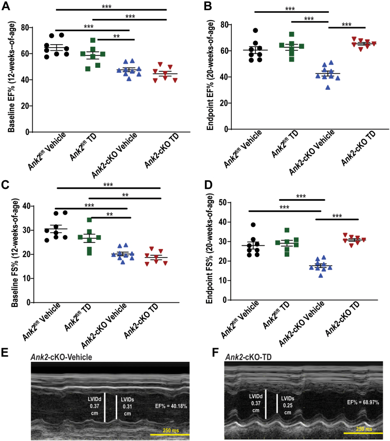
Arrhythmogenic cardiomyopathy (ACM) is an inherited heart disease, and current pharmacological therapies are directed toward the management of electrical manifestations. To date, none address the underlying pathophysiology of this progressive condition. We evaluated the therapeutic efficacy of Tideglusib (TD) in Ank2 cardio-selective-knockout and homozygous desmoglein-2 mutant ACM mouse models. TD was able to prevent and reverse the reduced cardiac function in treated mice. Moreover, TD-treated adult mice displayed a reduction in ventricular arrhythmia following adrenergic stimulation. We provide compelling preclinical data for TD as a potential therapy for patients with ACM.

Keywords: GSK-3β; Tideglusib; ankyrin-B; arrhythmia; arrhythmogenic cardiomyopathy; cardiovascular disease; desmoglein-2.


Conflict of interest statement

Funding Support and Author Disclosures The authors are supported by National Institutes of Health grants HL146969 and HL175380 to Dr Refaey; HL135754 to Dr Mohler; HL171689, HL156652, and HL165751 to Dr Hund; a grant from the Ohio State Frick Center for Heart Failure and Arrhythmia, the Leducq Foundation (TNE FANTASY 19CV03) to Dr Mohler; and the JB Cardiovascular Fellowship. Dr Roberts has been named as an inventor on a patent application that has been filed for use of Tideglusib as a therapeutic for ACM. All other authors have reported that they have no relationships relevant to the contents of this paper to disclose.

Figures

None
Graphical abstract
Figure 1
Figure 1
Tideglusib Prevents Cardiac Dysfunction Caused by AnkB Deficiency in Ank2-cKO Mice (A) Diagrammatic illustration of the structure of the study including the preventative study (4 to 12 weeks of age) and the reversal study (12 to 20 weeks of age), and the 4 groups involved in each study. (B) Percent ejection fraction (%EF) at 4 weeks of age before the prevention study, and before the onset of the phenotype associated with ankyrin-B deficiency, and (C) at 12 weeks of age after 8 weeks of Tideglusib (TD) or vehicle therapy. (D) Percent fractional shortening (%FS) at 4 weeks of age (before the prevention study) and (E) at 12 weeks of age at the conclusion of the prevention study. (F and G) Representative short-axis M-mode echocardiographic images at the conclusion of the prevention study of vehicle-treated Ank2 cardioselective knockout (Ank2-cKO) mice and TD-treated Ank2-cKO mice. Data presented using mean ± SEM and compared using Kruskal-Wallis with multiple comparisons test, n (Ank2fl/fl) = 7 (vehicle) and 8 (TD) and n (Ank2-cKO) = 7 (vehicle) and 8 (TD), ∗P < 0.05, ∗∗P < 0.01 and ∗∗∗P < 0.001.
Figure 2
Figure 2
Tideglusib Prevents the Progression of Cardiac Dysfunction in Dsg2mut/mut Mice (A) Percent ejection fraction (%EF) at 8 weeks and every 2 weeks till 16 weeks of age between vehicle-and drug-treated Dsg2mut/mut mice. (B) Heart rate (HR) between 8 and 16 weeks throughout the course of TD administration. (C) Left ventricular internal diameter at end-diastole (LVIDd) at baseline and following TD throughout the treatment period. (D) Left ventricular posterior wall at end diastole (LVPWd) every 2 weeks starting at 8 weeks comparing vehicle- and TD-treated Dsg2mut/mut mice. (E and F) Representative short-axis M-mode echocardiographic images at the conclusion of the prevention study of vehicle-treated Dsg2mut/mut and TD-treated Dsg2mut/mut mice. Data presented using mean ± SEM and compared using repeated measures analysis of variance , n = 9-11/treatment, ∗P < 0.05, ∗∗P < 0.01, and ∗∗∗P < 0.001.
Figure 3
Figure 3
Tideglusib Prevents Arrhythmia Generation in Ank2-cKO Mice (A) Representative conscious electrocardiogram recording captured via implantable telemeter device following concomitant administration of 120 mg/kg caffeine and 2mg/kg epinephrine via IP injection in Ank2fl/fl-vehicle-treated mouse, (B) Ank2-cKO-TD-treated mouse, (C) and Ank2-cKO-vehicle-treated mouse showing ventricular tachycardia (VT), (D) nonsustained ventricular tachycardia (NSVT), and (E) premature ventricular contractions (PVCs) with red arrows to highlight region of interest. (F) PVCs were quantified over 30 minutes immediately following injection. (G) Percentage of mice to experience at a minimum 1 run of NSVT and (H) VT are displayed. (I) Time in VT is displayed. Data presented using mean ± SEM and compared using Kruskal-Wallis test with Dunn’s multiple comparison test, n (Ank2fl/fl) = 3 (vehicle) and 4 (TD) and n (Ank2-cKO) = 6 (vehicle) and 5 (TD), ∗P < 0.05.
Figure 4
Figure 4
Tideglusib Reduces Arrhythmic Burden in Dsg2mut/mut Mice Following Epinephrine Stimulation (A) Representative conscious electrocardiogram recording captured via implantable telemeter device following 2 mg/kg epinephrine administration in Dsg2mut/mut treated mice. Number of (B) PVCs, (C) NSVT, (D) Couplet, and (E) VT throughout the recording time. (F) Percentage of mice that displayed NSVT and VT following adrenergic challenge. Data presented using mean ± SEM and compared using Mann-Whitney test (B, C, D and E), n = 4-5/treatment, ∗P < 0.05, and ∗∗P < 0.01. Abbreviations as in Figure 3.
Figure 5
Figure 5
Expression of AnkB, Total β-Catenin and Phosphorylated β-Catenin After Tideglusib Administration in Ank2-cKO Mice (A) Validation of the Ank2-cKO murine model via immunoblotting, and (B) quantitative analysis of AnkB protein levels in hearts of both Ank2fl/fl and Ank2-cKO mice. (C-D) Immunoblots and quantitative analysis of total β-catenin and GAPDH in Ank2-cKO-TD- vs vehicle-treated mice. (E-F) Immunoblots and quantitative analysis of phosphorylated β-catenin and GAPDH in Ank2-cKO TD- vs vehicle-treated mice. Data presented using mean ± SEM and compared using analysis of variance with Tukey post hoc test or Student's t-test, n = 3-4, ∗∗P < 0.01. Abbreviations as in Figures 1 and 3.
Figure 6
Figure 6
Effects of Tideglusib on GSK-3β Phosphorylation and β-Catenin Phosphorylation in Dsg2mut/mut Mice Immunoblots and quantitative analysis of (A) total GSK-3β, (B) phosphorylated form of GSK-3β, (C) total β-catenin and (D) phosphorylated form of β-catenin. Data presented using mean ± SEM and compared using Student's t-test (n = 8-9/group), ∗∗P < 0.01.
Figure 7
Figure 7
Tideglusib Reverses the Functional Phenotype in Ank2-cKO Mice (A) Percent ejection fraction (%EF) at 12 weeks of age before the reversal study, and after the onset of the phenotype associated with Ank2-cKO, and (B) at 20 weeks of age after 8 weeks of TD/vehicle therapy. (C) Percent fractional (%FS) shortening at 12 weeks of age (before the reversal study) and (D) at 20 weeks of age at the conclusion of the reversal study. (E and F) Representative short-axis M-echocardiographic images at the conclusion of the reversal study in Ank2-cKO vehicle-treated mice and Ank2-cKO TD-treated mice. Data presented using mean ± SEM and compared using analysis of variance with Tukey's post hoc test, n (Ank2fl/fl) = 8 (vehicle) and 7 (TD) and n (Ank2-cKO) = 9 (vehicle) and 7 (TD), ∗∗P < 0.01 and ∗∗∗P < 0.001. Abbreviations as in Figure 1.
Figure 8
Figure 8
Tideglusib Rescues the Arrhythmic Burden in Ank2-cKO Mice Following Epinephrine and Caffeine Stimulation (A) Representative conscious electrocardiogram recording captured via implantable telemeter devices following concomitant administration of 120 mg/kg caffeine and 2 mg/kg epinephrine via IP injection in Ank2fl/fl vehicle-treated mouse, (B) Ank2-cKO TD-treated mouse, (C) and Ank2-cKO vehicle-treated mouse showing a PVC, (D) NSVT, and (E) multiple runs of triplets/NSVT with red arrows to highlight region of interest. (F) PVCs/couplets and (G) runs of NSVT quantified per hour are displayed. (H) Percentage of mice to experience at a minimum one run of VT are displayed. Data presented using mean ± SEM and compared using Kruskal-Wallis test with Dunn’s multiple comparison test, n (Ank2fl/fl) = 5 (vehicle) and 4 (TD) and n (Ank2-cKO) =5 (vehicle) and 6 (TD), ∗∗P < 0.01 and ∗∗∗P < 0.001. Abbreviations as in Figures 1 and 3.
Figure 9
Figure 9
Tideglusib Restores the ID Distribution of JUP and Cx-43 in Dsg2mut/mut Mice (A and B) Representative heart cryosections and quantitative analysis from Dsg2mut/mut mice (treated with vehicle or TD) immuno-stained with N-cadherin (red), JUP (green) and overlap including DAPI nuclear stain (blue). Scale bars equal 20um. Images are representative of n (mouse) = 6 and n (intercalated disc [ID]) = 60 for vehicle and n (mouse) = 7 and n (ID) 70 for TD. Data did not pass Shapiro-Wilk normality test and Mann-Whitney U test was performed for statistical analysis. (C and D) Representative heart cryosections and quantitative analysis from Dsg2mut/mut mice (treated with vehicle or TD) immuno-stained with N-Cadherin (red), Cx-43 (green) and overlap including DAPI nuclear stain (blue). Scale bars equal 20um. Images are representative of n (mouse) = 5 and n (ID) = 50 for vehicle and TD. Data presented using mean ± SEM and compared using Student's t-test, ∗∗∗P < 0.001. Abbreviations as in Figure 1.

References

    1. Stevens T.L., Wallace M.J., Refaey M.E., Roberts J.D., Koenig S.N., Mohler P.J. Arrhythmogenic cardiomyopathy: molecular insights for improved therapeutic design. J Cardiovasc Dev Dis. 2020;7(2):21. - PMC - PubMed
    1. Frank R., Fontaine G., Vedel J., et al. [Electrocardiology of 4 cases of right ventricular dysplasia inducing arrhythmia] Archives des maladies du coeur et des vaisseaux. 1978;71:963–972. - PubMed
    1. Dungan W.T., Garson A., Jr., Gillette P.C. Arrhythmogenic right ventricular dysplasia: a cause of ventricular tachycardia in children with apparently normal hearts. Am Heart J. 1981;102:745–750. - PubMed
    1. Marcus F.I., McKenna W.J., Sherrill D., et al. Diagnosis of arrhythmogenic right ventricular cardiomyopathy/dysplasia proposed modification of the task force criteria. Circulation. 2010;121 1533-U118. - PMC - PubMed
    1. Corrado D., Perazzolo Marra M., Zorzi A., et al. Diagnosis of arrhythmogenic cardiomyopathy: the Padua criteria. Int J Cardiol. 2020;319:106–114. - PubMed

LinkOut - more resources