. 2025 Aug 27;15(1):31643.
doi: 10.1038/s41598-025-16908-3.

RAD18 methylation by the methyltransferase SETD6 attenuates DNA breaks

Affiliations

RAD18 methylation by the methyltransferase SETD6 attenuates DNA breaks

Lital Estrella Weil et al. Sci Rep. .

Abstract

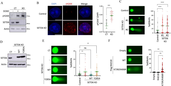
This study investigated the interaction between the SETD6 lysine methyltransferase and RAD18, a key protein in the DNA damage repair pathway. SETD6 belongs to the SET-domain-containing family of proteins, which are known to catalyze protein methylation, a post-translational modification that plays a critical role in regulating protein function, stability, and interactions. Using protein microarray technology, we identified RAD18 as an interactor and substrate of SETD6. We confirmed this interaction through ELISA and immunoprecipitation assays, demonstrating that SETD6 directly binds and methylates RAD18. Using mass spectrometry and site-directed mutagenesis, we identified that RAD18 undergoes mono-methylation at the K73 and K406 residues. Furthermore, we found that RAD18 methylation affects its nuclear localization. Specifically, SETD6 KO cells exhibited increased nuclear RAD18 levels, suggesting that methylation status influences RAD18's shuttling between the cytoplasm and nucleus. Notably, depletion of SETD6 led to elevated markers of DNA damage (γH2AX) and increased DNA breaks, as evidenced by comet assays. Restoring SETD6 activity significantly reduced DNA damage, while a catalytic inactive mutant did not have this effect, underscoring the importance of SETD6's enzymatic function. Overall, our results demonstrate that SETD6-mediated methylation of RAD18 is essential for attenuating DNA breaks, thereby regulating its cellular localization and function in maintaining genomic integrity.


Conflict of interest statement

Declarations. Competing interests: The authors declare no competing interests.

Figures

Fig. 1
Fig. 1
SETD6 binds RAD18 in-vitro and in cells. (A) Schematic representation of the M-NAPPA process. DNA sequences encoding specific proteins were spotted onto a glass slide. The DNA is then expressed in-situ using a cell-free expression system, creating a protein microarray. The interaction was detected using fluorescence following data analysis. (B) Enzyme-linked immunosorbent assay (ELISA) was performed with the indicated recombinant proteins. Self-interaction between His-SETD6 and GST-SETD6 served as a positive control. The graph represents absorbance at 450 nm for each condition. ****p-value < 0.0001. (C) HeLa whole cell lysates were immunoprecipitated with agarose beads conjugated to the indicated antibody, followed by Western blot analysis with indicated antibodies.
Fig. 2
Fig. 2
SETD6 methylates RAD18 on K73 and K406. (A) In-vitro methylation assay in the presence of 3H-labeled SAM with recombinant His-SETD6 and His-RAD18. The methylated proteins were detected by autoradiogram and a Coomassie stain of the recombinant proteins used in the reactions is shown on the bottom. (B) Semi-in-vitro methylation assay of immunoprecipitated Flag-RAD18 that was over-expressed in PC3 cells, with and without the presence of His-SETD6. The methylated proteins were detected by autoradiogram and Flag-RAD18 was visualized using Western blot analysis using αFlag antibody. (C) HeLa cells lysates were immunoprecipitated with Pan-Methyl lysine antibody followed by Western Blot analysis with the indicated antibodies. (D) MS spectra of RAD18 TQCPTCCVTVTEPDLK73NNR and LSSVCMGQEDNMTSVTNHFSQSK406. Major y- and b-ions are displayed as red, and blue, respectively. (E) In-vitro methylation assay in the presence of 3H-labeled SAM with the indicated recombinant proteins. Coomassie stain of the recombinant proteins used in the reactions is shown on the bottom.
Fig. 3
Fig. 3
RAD18 is differentially recruited to the nucleus based on its methylation state. (A) Control and SETD6 KO cells were stained for DNA (DAPI) and endogenous RAD18, as indicated. Total integrated density of RAD18 in the nuclei was analyzed using Fiji software. (B) HeLa cells overexpressing Flag-RAD18 WT or K73R/K406R were stained for DNA (DAPI) and Flag, as indicated. Total integrated density was analyzed as in A. Graph values represent mean ± SEM. **p-value < 0.01 ****p-value < 0.0001. Scale, 10µM.
Fig. 4
Fig. 4
RAD18 methylation is linked to DNA damage accumulation. (A) Control and SETD6 KO cells were subjected to Western blot analysis to evaluate γH2AX levels. Control cells pre-treated with Doxorubicin served as positive control. Actin served as loading control. (B) Control and SETD6 KO HeLa cells were stained for endogenous γH2AX, as indicated. Total integrated density of γH2AX was analyzed using Fiji software. Graph values represent mean ± SEM. ****p-value < 0.0001. (C) Neutral comet assay in control and SETD6 KO cells is presented with representative Images taken with Evos fluorescence microscope and analysis was performed using CaspLab software. Graph values represent mean tail DNA%. (D) Western blot analysis with the indicated antibodies for CT and SETD6 KO cells rescued with SETD6 WT or SETD6 Y285A catalytic inactive mutant (E) HeLa control, SETD6 KO and SETD6 KO cells rescued with Flag-SETD6 and SETD6 Y285A catalytic inactive mutant and were treated as in C. Graph values represent mean ± SEM. *p-value < 0.05 **p-value < 0.01 ****p-value < 0.0001. Scale, 10µM. (F) Cells overexpressing RAD18 WT or K73R, K406R were subjected to Neutral comet assay as in C. Graph values represent mean ± SEM ****p-value < 0.0001. Scale, 10µM.

Similar articles

References

    1. Alvarez-Venegas, R. & Avramova, Z. SET-domain proteins of the Su(var)3–9, E(z) and trithorax families. Gene285, 25–37 (2002). - PubMed
    1. Arrowsmith, C. H., Bountra, C., Fish, P. V., Lee, K. & Schapira, M. Epigenetic protein families: a new frontier for drug discovery. Nat. Rev. Drug Discov. 11, 384–400 (2012). - PubMed
    1. Levy, D. Lysine methylation signaling of non-histone proteins in the nucleus. Cell. Mol. Life Sci.76, 2873–2883 (2019). - PMC - PubMed
    1. Chopra, A., Feldman, M. & Levy, D. Orchestrating epigenetics: a comprehensive review of the methyltransferase SETD6. Exp. Mol. Med.57, 533–544 (2025). - PMC - PubMed
    1. Mukherjee, N., Cardenas, E., Bedolla, R. & Ghosh, R. SETD6 regulates NF-kappaB signaling in urothelial cell survival: implications for bladder cancer. Oncotarget8, 15114–15125 (2017). - PMC - PubMed

LinkOut - more resources