. 2025 Aug 27;25(1):91.
doi: 10.1186/s12896-025-01027-8.

Anticancer effects of folic acid-functionalized covalent organic framework containing doxorubicin on SW480 colon cancer cells: a promising tool for drug targeted delivery

Affiliations

Anticancer effects of folic acid-functionalized covalent organic framework containing doxorubicin on SW480 colon cancer cells: a promising tool for drug targeted delivery

Razie Ezati et al. BMC Biotechnol. .

Abstract

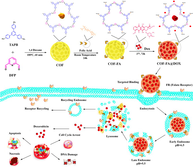
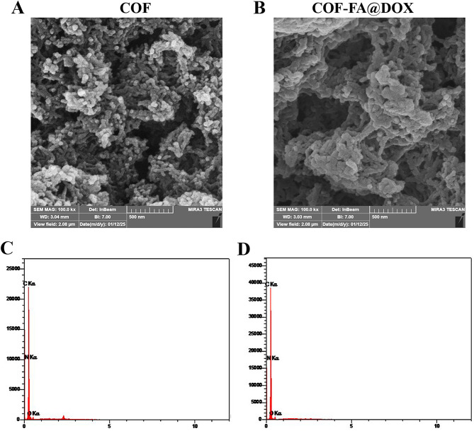
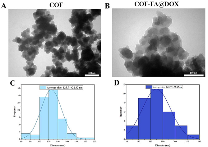
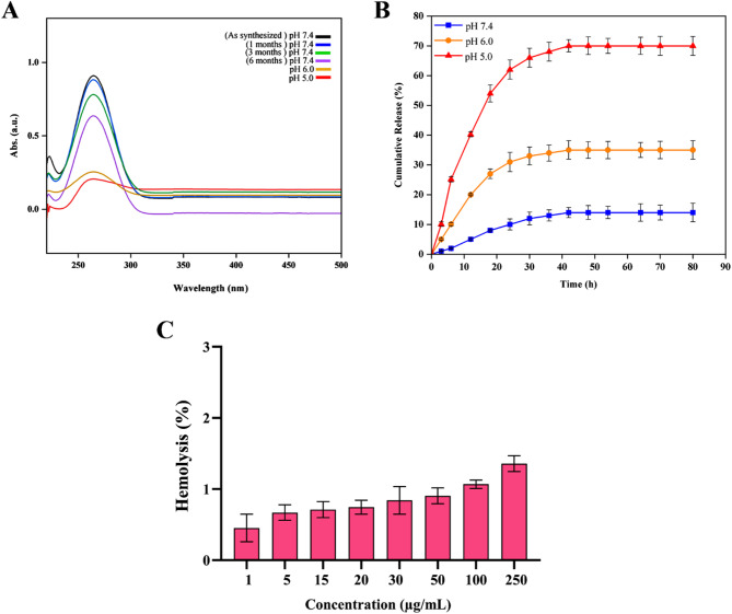
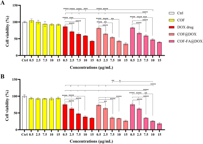
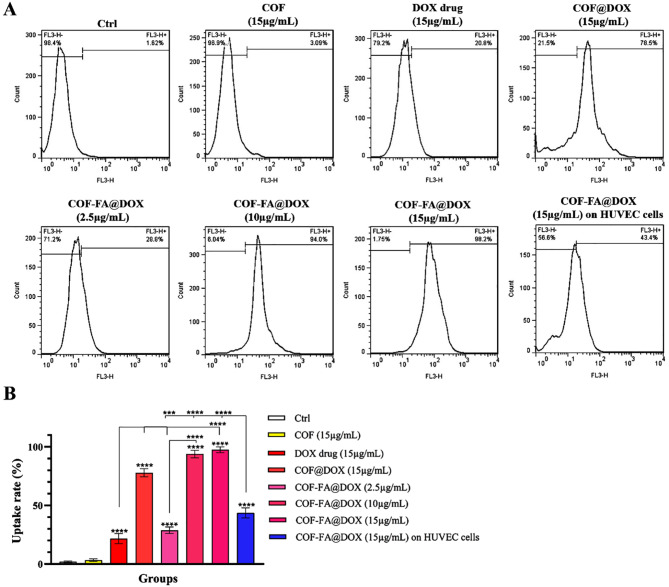
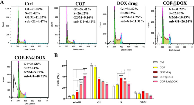
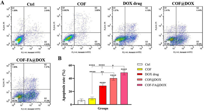
Colorectal cancer is one of the deadliest forms of gastrointestinal cancer, with conventional treatments often facing significant limitations. As a result, new approaches, particularly in targeted drug delivery, have shown great promise. In this study, the COF-FA@DOX nanocarrier was developed, where covalent organic frameworks (COFs) were functionalized with folic acid (FA) and then loaded with Doxorubicin (DOX). The as-synthesized COF-FA@DOX nanocarrier was characterized using different techniques. To assess its anticancer effectiveness, MTT, flow cytometry, and scratch assays were conducted on SW480 and HUVEC cells to examine cell viability, cellular uptake, cell cycle progression, apoptosis, and cell migration, respectively. The obtained results demonstrated that the COF-FA@DOX nanocarrier was efficiently internalized by cancer cells and showed significantly higher cytotoxicity compared to other synthesized nanocarrier groups and free DOX drug. Moreover, the COF-FA@DOX nanocarrier caused cell cycle arrest, induced apoptosis, and inhibited cell migration at lower doses than the free DOX drug. Altogether, these findings suggest that the COF-FA@DOX nanocarrier is an effective and promising drug delivery system for DOX in colorectal cancer, potentially enhancing the therapeutic efficacy of DOX drug while minimizing side effects through targeted delivery. Further investigation is required to assess their efficacy in vivo and discover potential clinical applications.

Keywords: Colorectal cancer; Covalent organic frameworks; Doxorubicin; Drug delivery; Folic acid; Targeted therapy.


Conflict of interest statement

Declarations. Ethics approval and consent to participate: Not applicable. Consent for publication: Not applicable. Competing interests: The authors declare no competing interests. Declaration of competing interest: The authors declare that they have no known competing financial interests or personal relationships that could have appeared to influence the work reported in this paper.

Figures

Fig. 1
Fig. 1
Schematic representation of the COF synthesis (yellow powder) and its subsequent functionalization with folic acid, followed by doxorubicin loading (red powder) and anticancer effects of COF-FA@DOX on colon cancer cells. All chemical structures were drawn using ChemBioOffice Ultra 2014
Fig. 2
Fig. 2
(A) FTIR spectra of the synthesized COF, along with its building blocks TAPB and DFP. (B) PXRD pattern of the COF, showing defined diffraction peaks that indicate the formation of a crystalline structure. (C) Nitrogen adsorption–desorption isotherms of the COF measured at 77 K, displaying a type II isotherm profile. (D) Pore size distribution calculated using the BJH method
Fig. 3
Fig. 3
High-resolution XPS analysis of the synthesized COF. (A) XPS survey spectrum illustrating the overall elemental composition of the COF, confirming the presence of carbon, nitrogen, and oxygen. (B) High-resolution C 1s spectrum, with deconvoluted peaks assigned to C = C, C–C, C–N = C, and C–O groups (C) O 1s spectrum showing deconvoluted peaks corresponding to O = C and O–C. (D) N 1s spectrum, revealing components associated with imine-type and pyridinic nitrogen species, further supporting the imine-linked COF structure
Fig. 4
Fig. 4
(A) FTIR spectra of COF functionalized with folic acid (COF-FA). (B) FTIR spectra of the DOX-loaded COF-FA nanocarrier (COF-FA@DOX). (C) Particle size distribution of COF, COF-FA, and COF-FA@DOX as measured by dynamic light scattering analysis. (D) Zeta potential of COF, COF-FA, and COF-FA@DOX nanocarrier
Fig. 5
Fig. 5
Field emission scanning electron microscopy (FESEM) image of the COF, showing its surface morphology (A). FESEM image of the DOX-loaded, folic acid-functionalized COF nanocarrier (B). Energy-dispersive X-ray spectroscopy (EDX) analysis of the COF confirms the elemental composition of the synthesized framework (C). EDX spectrum of the COF-FA@DOX nanocarrier (D)
Fig. 6
Fig. 6
Transmission electron microscopy (TEM) image of the synthesized COF (A). TEM image of the COF-FA@DOX nanocarrier (B). Particle size distribution of the COF, calculated based on TEM image analysis, indicating uniform nanoscale dimensions (C). Particle size distribution of the COF-FA@DOX nanocarrier derived from TEM analysis, reflecting a slight size increase after functionalization and drug encapsulation (D)
Fig. 7
Fig. 7
(A) Stability study of the synthesized COF at physiological and acidic pH values (7.4, 6.0, and 5.0). (B) DOX release profile from COF-FA@DOX nanocarrier under different pH conditions (pH 7.4, 6.0, and 5.0), simulating physiological and tumor-like environments. Data are presented as mean ± SD (n = 3). Error bars represent standard deviation. (C) Hemolysis percentage of red blood cells incubated with various concentrations of COF-FA@DOX nanocarrier, indicating acceptable hemocompatibility
Fig. 8
Fig. 8
Cell viability evaluation of various nanostructure formulations (COF, COF@DOX, and COF-FA@DOX nanocarriers) and free DOX across a concentration range of 0.5–15 µg/mL on SW480 cells at (A) 24 and (B) 48 h using MTT assay. Results are expressed as mean ± SD (n = 3), and statistical analysis was performed: *p < 0.05, **p < 0.01, ***p < 0.001, ****p < 0.0001
Fig. 9
Fig. 9
Cellular uptake assay of different formulations in SW480 (highly expressed folate receptors) and HUVEC (Low expressed folate receptors) cells. (A) Flow cytometry histograms showing treatment groups of different nanocarriers at various concentrations. (B) Quantitative analysis of cellular uptake rates (%) for each treatment group. Data are presented as mean ± SD (n = 3). Statistical significance: ***p < 0.001, ****p < 0.0001
Fig. 10
Fig. 10
Cell cycle evaluation in SW480 cells treated with various nanostructure formulations (COF, COF@DOX, and COF-FA@DOX nanocarriers) and free DOX drug concentration of 7.5 µg/mL for 48 h. (A) Flow cytometry histograms show the percentage of cells in the G1, S, G2/M, and sub-G1 phases. (B) Quantitative comparison of cell cycle distribution percentages among treated groups. The sub-G1 population indicates apoptotic cells. Results are presented as mean ± SD (n = 3). Statistical significance is marked as *p < 0.05, **p < 0.01, ****p < 0.0001
Fig. 11
Fig. 11
Apoptosis evaluation in SW480 cells treated with various nanostructure formulations (COF, COF@DOX, and COF-FA@DOX nanocarriers) and free DOX drug concentration of 7.5 µg/mL for 48 h. (A) Flow cytometry scatter plots illustrating cell distribution in Q1, Q2, Q3, and Q4 represent necrotic cells, late apoptotic cells, early apoptotic cells, and live cells, respectively. (B) Quantitative comparison of the apoptosis rate (%) among treated groups, calculated as the total percentage of early and late apoptotic cells (Q2 + Q3) for each group. Data are expressed as mean ± SD (n = 3). Statistical significance is indicated by *p < 0.05, **p < 0.01, ****p < 0.0001
Fig. 12
Fig. 12
(A) Cell migration inhibition evaluation in SW480 cells in 0 h and after 96 h of treatment with various nanostructure formulations (COF NPs, COF@DOX, and COF-FA@DOX nanocarriers) and free DOX drug concentration of 2.5 µg/mL using scratch assay. (B) Quantitative comparison of the cell migration inhibition (%) among treated groups. Data are presented as mean ± SD (n = 3). Statistical significance: **p < 0.05, ***p < 0.001, ****p < 0.0001

Similar articles

References

    1. Roshandel G, Ghasemi-Kebria F, Malekzadeh R. Colorectal cancer: epidemiology, risk factors, and prevention. Cancers. 2024;16(8):1530. - PMC - PubMed
    1. Hossain MS, Karuniawati H, Jairoun AA, Urbi Z, Ooi DJ, John A, et al. Colorectal cancer: A review of carcinogenesis, global epidemiology, current challenges, risk factors, preventive and treatment strategies. Cancers. 2022;14(7):1732. - PMC - PubMed
    1. Kuipers EJ, Rösch T, Bretthauer M. Colorectal cancer screening–optimizing current strategies and new directions. Nat Rev Clin Oncol. 2013;10(3):130–42. - PubMed
    1. Cai S, Thati S, Bagby TR, Diab HM, Davies NM, Cohen MS, et al. Localized doxorubicin chemotherapy with a biopolymeric nanocarrier improves survival and reduces toxicity in xenografts of human breast cancer. J Control Release. 2010;146(2):212–8. - PMC - PubMed
    1. Sazonova EV, Yapryntseva MA, Pervushin NV, Tsvetcov RI, Zhivotovsky B, Kopeina GS. Cancer drug resistance: targeting proliferation or programmed cell death. Cells. 2024;13(5):388. - PMC - PubMed

MeSH terms