Review
. 2025 Aug 19;15(8):884.
doi: 10.3390/brainsci15080884.

Redox-Regulated Pathways in Glioblastoma Stem-like Cells: Mechanistic Insights and Therapeutic Implications

Affiliations
Review

Redox-Regulated Pathways in Glioblastoma Stem-like Cells: Mechanistic Insights and Therapeutic Implications

Nadia Fernanda Esteban-Román et al. Brain Sci. .

Abstract

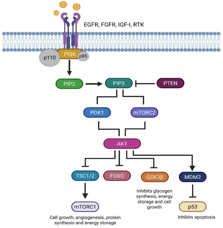
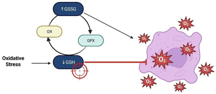
Glioblastoma (GBM) is the most aggressive primary brain tumor, characterized by rapid proliferation, invasiveness, therapeutic resistance, and an immunosuppressive tumor microenvironment. A subpopulation of glial stem-like cells (GSCs) within GBM tumors contributes significantly to tumor initiation, progression, and relapse, displaying remarkable adaptability to oxidative stress and metabolic reprogramming. Recent evidence implicates the atypical kinases RIOK1 and RIOK2 in promoting GBM growth and proliferation through their interaction with oncogenic pathways such as AKT and c-Myc. Concurrently, the redox-sensitive Nrf2/Keap1 axis regulates antioxidant defenses and supports GSC survival and chemoresistance. Additionally, aberrant activation of the canonical Wnt/β-catenin pathway in GSCs enhances their self-renewal, immune evasion, and resistance to standard therapies, particularly under oxidative stress conditions. This review integrates current knowledge on how redox homeostasis and key signaling pathways converge to sustain GSC maintenance and GBM malignancy. Finally, we discuss emerging redox-based therapeutic strategies designed to target GSC resilience, modulate the tumor immune microenvironment, and surmount treatment resistance.

Keywords: cancer stem cells; glial stem-like cells; glioblastoma; glioma; oxidative stress; redox-targeted therapy.


Conflict of interest statement

The authors declare no conflicts of interest.

Figures

Figure 1
Figure 1
Mitochondrial enzyme aldehyde dehydrogenase 1 family member 2 (ALDH1L2) catalyzes the conversion of 10-formyl-tetrahydrofolate (10-formyl-THF) into carbon dioxide (CO2) and tetrahydrofolate (THF), generating reduced nicotinamide adenine dinucleotide phosphate (NADPH). NADPH serves as a cofactor for the enzyme glutathione reductase (GSH reductase), which reduces oxidized glutathione (GSSG) to its active form, reduced glutathione (GSH). Glutathione peroxidase (GPX) then utilizes GSH to convert peroxides into water or alcohol, thereby contributing to cellular redox homeostasis (created with https://BioRender.com). The ALDH1L2 enzyme is crucial for redox balance in glioma stem-like cells (GSCs). By synthesizing NADPH, ALDH1L2 enhances the glutathione antioxidant system, which allows GSCs to protect themselves from oxidative stress and survive in the tumor microenvironment. ALDH1L2 dysfunction compromises antioxidant defenses, increasing intracellular ROS levels and reducing the aggressive phenotype of GSCs. Therefore, ALDH activity not only preserves redox balance and mitochondrial function but also serves as a biomarker for tumor aggressiveness and therapy resistance. This mechanism highlights the importance of redox signaling pathways in GSC maintenance and aggressiveness.
Figure 2
Figure 2
CD44 depletion in glioblastoma cells enhances their response to oxidative stress induced by antitumor agents such as temozolomide (TMZ) and carmustine (BCNU). Reactive oxygen species (ROS) activate the Hippo pathway by phosphorylating the merlin protein, which, in turn, triggers the phosphorylation and activation of Mammalian Sterile Twenty-like kinases 1 and 2 (MST1/2) and Large Tumor Suppressors 1 and 2 (LATS1/2). This cascade leads to the phosphorylation and inactivation of Yes-associated protein (YAP), preventing its nuclear translocation. As a result, the expression of anti-apoptotic proteins cIAP1/2 is inhibited, while levels of cleaved caspase-3 increase, ultimately promoting apoptosis in tumor cells (created with https://BioRender.com).
Figure 3
Figure 3
FTRAP1 (HSP75) modulates mitochondrial metabolism by suppressing succinate dehydrogenase (SDH), diminishing the production of reactive oxygen species (ROS), and stabilizing HIF1α. This facilitates metabolic reprogramming towards aerobic glycolysis, enhancing the activation of glioma stem cells (GSCs). Nonetheless, TRAP1 can activate SDH in specific situations, enhancing mitochondrial respiration in GSCs, hence contributing to their metabolic flexibility and treatment resistance (created with https://BioRender.com).
Figure 4
Figure 4
The figure illustrates the role of Sirtuin 3 (SIRT3) as a mitochondrial regulator of redox equilibrium and cellular oxidative stress. Its function in activating antioxidant enzymes such as MnSOD and IDH, modulating ROS, boosts cell survival, respiration, and tumor microenvironment efficiency (created with https://BioRender.com).
Figure 5
Figure 5
Schematic representation of the bidirectional TRAP1-SIRT3 functional axis in glioma stem cells (GSCs). TRAP1 augments the enzymatic function of SIRT3, facilitating the deacetylation and activation of SOD2, hence diminishing mitochondrial reactive oxygen species (ROS) levels. SIRT3 modulates the acetylation of TRAP1, creating a positive feedback loop between the two proteins. This functional axis enhances mitochondrial respiration efficiency by modulating electron transport (ETC), particularly in low glucose availability situations. The interplay between TRAP1 and SIRT3 enhances GSC metabolic adaptability under stress, hence augmenting their survival, self-renewal ability, and respiratory efficiency in challenging tumor microenvironments (created with https://BioRender.com).
Figure 6
Figure 6
Regulation of Nrf2 under physiological and oxidative stress conditions. (A) Under normal physiological conditions, Keap1 binds Nrf2 through cysteine residues (Cys151, Cys273, and Cys288), facilitating its ubiquitination via the Cul3-ROC1 E3 ligase complex, leading to proteasomal degradation. (B) In an oxidative microenvironment, reactive oxygen species (ROS) modify the cysteine residues on Keap1, inhibiting its ability to promote Nrf2 ubiquitination. As a result, stabilized Nrf2 accumulates and translocates into the nucleus, where it forms a heterodimer with sMaf proteins and binds to antioxidant response elements (AREs), activating the transcription of antioxidant and metabolic genes (created with https://BioRender.com).
Figure 7
Figure 7
Interaction between Wnt/β-catenin signaling and reactive oxygen species (ROS) in the regulation of cellular functions. Activation of Wnt ligands binds Frizzled and LRP receptors, leading to Dishevelled activation and subsequent inhibition of the β-catenin destruction complex (GSK-3β, Axin, CKIα, and APC). Stabilized β-catenin translocates into the nucleus, where it interacts with TCF/LEF and FOXO transcription factors to promote the expression of target genes such as c-myc and Cyclin D1, supporting stem cell maintenance, proliferation, and invasion. Concurrently, Wnt signaling activates PI3K-Akt, which inhibits TSC1/2, enabling Rheb-mediated activation of mTORC1, thereby promoting cell proliferation and autophagy. ROS, produced by NOX-1, enhance Wnt signaling by inhibiting NRX, a negative regulator of Dishevelled, further amplifying β-catenin signaling (created with https://BioRender.com).
Figure 8
Figure 8
Degradation of oxidatively modified GAPDH under oxidative stress. Hydrogen peroxide (H2O2) exposure leads to oxidative modification of glyceraldehyde-3-phosphate dehydrogenase (GAPDH), contributing to decreased protein levels and enzymatic activity. Likewise, a major modification is by 4-hydroxy-2-nonenal (4-HNE), a cytotoxic lipid peroxidation byproduct that covalently binds to GAPDH. Cathepsin G, a serine protease featuring a catalytic triad (Asp-His-Ser), is implicated in the degradation of 4-HNE-modified GAPDH (created with https://BioRender.com).
Figure 9
Figure 9
Glutathione redox cycling in the oxidative tumor microenvironment. Elevated oxidative stress in the tumor microenvironment (TME) leads to depletion of reduced glutathione (GSH), resulting in accumulation of its oxidized form (GSSG). Glutathione peroxidase (GPX) catalyzes the reduction of reactive oxygen species (ROS) using GSH, while glutathione reductase (GR) regenerates GSH from GSSG to maintain redox homeostasis. Excessive ROS, if not neutralized, causes extensive damage to DNA, proteins, and lipids, promoting cellular dysfunction and death. Targeting the GSH system has emerged as a promising redox-sensitive therapeutic approach, especially in tumors with elevated GSH levels that confer resistance to oxidative damage (created with https://BioRender.com).

Similar articles

References

    1. Finch A., Solomou G., Wykes V., Pohl U., Bardella C., Watts C. Advances in research of adult gliomas. Int. J. Mol. Sci. 2021;22:924. doi: 10.3390/ijms22020924. - DOI - PMC - PubMed
    1. Baker S.J., Zong H., Monje M. Glial Malignancies. Cold Spring Harb. Perspect. Biol. 2024;17:a041373. doi: 10.1101/cshperspect.a041373. - DOI - PubMed
    1. Aiman W., Gasalberti D.P., Rayi A. StatPearls. StatPearls Publishing; St. Petersburg, FL, USA: 2025. Low-Grade Gliomas. - PubMed
    1. Louis D.N., Perry A., Wesseling P., Brat D.J., Cree I.A., Figarella-Branger D., Hawkins C., Ng H.K., Pfister S.M., Reifenberger G., et al. The 2021 WHO Classification of Tumors of the Central Nervous System: A summary. Neuro Oncology. 2021;23:1231–1251. doi: 10.1093/neuonc/noab106. - DOI - PMC - PubMed
    1. Lopez-Gonzalez M.A., Sotelo J. Brain tumors in Mexico: Characteristics and prognosis of glioblastoma. Surg. Neurol. 2000;53:157–162. doi: 10.1016/S0090-3019(99)00177-9. - DOI - PubMed

LinkOut - more resources