. 2025 Jul 23;15(8):1064.
doi: 10.3390/biom15081064.

A Distinct miRNA Profile in Intimal Hyperplasia of Failed Arteriovenous Fistulas Reveals Key Pathogenic Pathways

Affiliations

A Distinct miRNA Profile in Intimal Hyperplasia of Failed Arteriovenous Fistulas Reveals Key Pathogenic Pathways

Carmen Ciavarella et al. Biomolecules. .

Abstract

Intimal hyperplasia (IH) compromises the patency of arteriovenous fistula (AVF) vascular access in patients with end-stage kidney disease. Uncontrolled cell proliferation and migration, driven by inflammation, shear stress and surgery, are well-known triggers in IH. Recently, microRNAs (miRNAs) have emerged as regulators of core mechanisms in cardiovascular diseases and as potential markers of IH. This study was aimed at identifying a specific miRNA panel in failed AVFs and clarifying the miRNA involvement in IH. miRNA profiling performed in tissues from patients with IH (AVFs) and normal veins (NVs) highlighted a subset of four miRNAs significantly deregulated (hsa-miR-155-5p, hsa-miR-449a-5p, hsa-miR-29c-3p, hsa-miR-194-5p) between the two groups. These miRNAs were analyzed in tissue-derived cells (NVCs and AVFCs), human aortic smooth muscle cells (HAOSMCs) and human umbilical vein endothelial cells (HUVECs). The panel of hsa-miR-449a-5p, hsa-miR-155-5p, hsa-miR-29c-3p and hsa-miR-194-5p was up-regulated in AVFCs, HAOSMCs and HUVEC under inflammatory stimuli. Notably, overexpression of hsa-miR-449a-5p exacerbated the proliferative, migratory and inflammatory features of AVFCs. In vitro pharmacological modulation of these miRNAs with pioglitazone, particularly the down-regulation of hsa-miR-155-5p and hsa-miR-29c-3p, suggested their involvement in IH pathogenesis and a potential translational application. Overall, these findings provide new insights into the pathogenesis of AVF failure, reinforcing the miRNA contribution to IH detection and prevention.

Keywords: arteriovenous fistula failure; hsa-miR-449a-5p; intimal hyperplasia; miRNA; migration; proliferation.


Conflict of interest statement

The authors declare no conflicts of interest.

Figures

Figure 1
Figure 1
Expression profile of miRNAs in AVFs compared to normal veins. (a) Volcano plot representation of the miRNA profile in human AVFs compared with NV. Data are reported as fold changes (log2) versus p value (−log10). Green points: down-regulated miRNAs; red points: up-regulated miRNAs. Fold change boundary: 2; p-value: 0.05 (points above the horizontal line are miRNAs significantly deregulated). The graphic representation was obtained with Expression Suite Software v1. (b) Validation by RT-qPCR of selected miRNAs (hsa-miR-155-5p, hsa-miR-449a-5p, hsa-miR-29c-3p, hsa-miR-194-5p) in AVF veins compared to the NV group. The AVF group was further divided in two subgroups: AVF IH, carrying intimal hyperplasia; AVF aneurysm, with aneurysmal degeneration of the vascular wall. Small nuclear RNA U6 (snU6) was selected as an endogenous control, and the relative miRNA expression was calculated with the 2−∆∆CT method. Data are expressed as mean ± SD, and statistical analysis was performed by unpaired Student t test; * p < 0.05, ** p < 0.01. NV, normal veins; AVF, arteriovenous fistula; FC, fold changes.
Figure 2
Figure 2
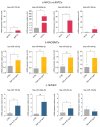
The miRNA expression pattern in human vascular cells mirrors the AVF tissue signature. RT-qPCR for hsa-miR-155-5p, hsa-miR-449a-5p, hsa-miR-29c-3p and hsa-miR-194b was performed in (a) AVFCs compared to NVCs, (b) HAOSMC and (c) HUVEC exposed to chronic inflammatory injury by TNF-α administration for 6 days (25 ng/mL) compared to untreated controls. Small nuclear RNA U6 (snU6) was selected as endogenous control, and the relative miRNA expression was calculated with the 2−∆∆CT method. Data are reported as mean ± SD of at least three independent experiments; * p < 0.05, ** p < 0.01, *** p < 0.001; unpaired Student t-test.
Figure 3
Figure 3
Pioglitazone modulates IH-associated miRNA expression in vascular cell models. Pioglitazone differently modulates the IH-miRNA expression levels in (a) AVFCs, (b) HAOSMCs and (c) HUVEC, by down-regulating the levels of hsa-miR-29c-3p and hsa-miR-155-5p and up-regulating hsa-miR-449a-5p in AVFCs. Results are expressed as fold changes relative to untreated controls. Small nuclear RNA U6 (snU6) was selected as endogenous control, and the relative miRNA expression was calculated with the 2−∆∆CT method. Data are reported as mean ± SD of at least three independent experiments; * p < 0.05, ** p < 0.01, unpaired Student t-test.
Figure 4
Figure 4
hsa-miR-449a-5p contributes to the main IH pathogenic mechanisms by stimulating the proliferative and migratory cell phenotype. (a) Validation of hsa-miR-449a-5p over-expression by transient transfection in NVCs and AVFCs after 48 h. Effects of hsa-miR-449a over-expression on NVCs and AVFCs’ proliferation, measured by (b) BrdU incorporation as a marker of newly synthesized DNA and (c) Ki-67 staining by immunofluorescence. For BrdU, absorbance values were analyzed at the 450 nm optical density (OD) with a reference wavelength of 595 nm by a Spark multimode microplate reader (Tecan). For Ki-67 expression, data are reported as the ratio of Ki-67 positive cell number to the total cell number. Green: Ki-67, blue: DAPI. Scale bar: 25 μm, 40× magnification. (d) Analysis of NVCs and (e) AVFCs’ migration performed through a scratch assay by using an Incucyte S3 instrument. The following migration parameters were evaluated: wound width, expressed in micrometers (μm), and wound confluence, expressed as a percentage (%). Representative images of wound healing taken with a 10× objective lens at the following time lapses: 0 h (initial wound process), 6 h, 33 h and 36 h for NVCs; 0 h, 12 h, 15 h and 18 h for AVFCs. Yellow: scratch wound width; pink: wound confluence (cells migrated into the scratch area). Data are reported as mean ± SD of at least three independent experiments. Statistical analysis was performed with an unpaired Student t test for (ac), and a two-way ordinary ANOVA test followed by Sidak’s multiple-comparisons tests for (d,e); * p < 0.05; ** p < 0.01. Abbreviations: BrdU, bromo-deoxy-uridine; OD, optical density; miR-NC, negative control; miR-mimic, hsa-miR-449a-5p over-expression; NVCs, normal vein cells; AVFCs, arteriovenous fistula cells.

References

    1. Ma S., Duan S., Liu Y., Wang H. Intimal Hyperplasia of Arteriovenous Fistula. Ann. Vasc. Surg. 2022;85:444–453. doi: 10.1016/j.avsg.2022.04.030. - DOI - PubMed
    1. Déglise S., Bechelli C., Allagnat F. Vascular Smooth Muscle Cells in Intimal Hyperplasia, an Update. Front. Physiol. 2023;13:1081881. doi: 10.3389/fphys.2022.1081881. - DOI - PMC - PubMed
    1. Roy-Chaudhury P., Arend L., Zhang J., Krishnamoorthy M., Wang Y., Banerjee R., Samaha A., Munda R. Neointimal Hyperplasia in Early Arteriovenous Fistula Failure. Am. J. Kidney Dis. 2007;50:782–790. doi: 10.1053/j.ajkd.2007.07.019. - DOI - PubMed
    1. Martinez L., Tabbara M., Duque J.C., Selman G., Falcon N.S., Paez A., Griswold A.J., Ramos-Echazabal G., Hernandez D.R., Velazquez O.C., et al. Transcriptomics of Human Arteriovenous Fistula Failure: Genes Associated With Nonmaturation. Am. J. Kidney Dis. 2019;74:73–81. doi: 10.1053/j.ajkd.2018.12.035. - DOI - PMC - PubMed
    1. Chakraborty R., Chatterjee P., Dave J.M., Ostriker A.C., Greif D.M., Rzucidlo E.M., Martin K.A. Targeting Smooth Muscle Cell Phenotypic Switching in Vascular Disease. JVS-Vasc. Sci. 2021;2:79–94. doi: 10.1016/j.jvssci.2021.04.001. - DOI - PMC - PubMed

LinkOut - more resources