. 2025 Aug 21;15(8):1204.
doi: 10.3390/biom15081204.

PInteract: Detecting Aromatic-Involving Motifs in Proteins and Protein-Nucleic Acid Complexes

Affiliations

PInteract: Detecting Aromatic-Involving Motifs in Proteins and Protein-Nucleic Acid Complexes

Dong Li et al. Biomolecules. .

Abstract

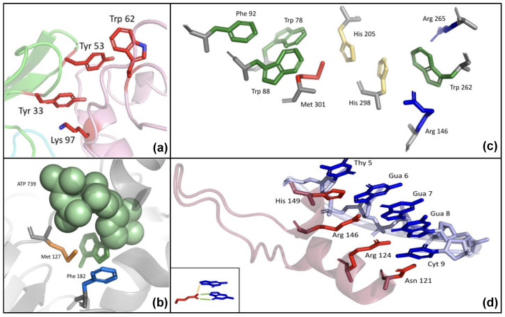
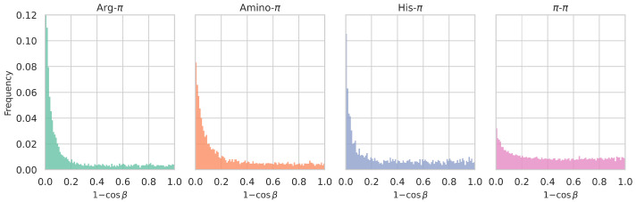
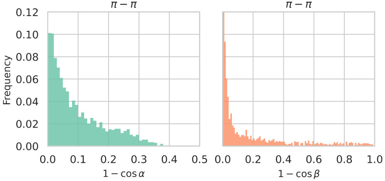
With the recent development of accurate protein structure prediction tools, virtually all protein sequences now have an experimental or a modeled structure. It has therefore become essential to develop fast algorithms capable of detecting non-covalent interactions not only within proteins but also in protein-protein, protein-DNA, protein-RNA, and protein-ligand complexes. Interactions involving aromatic compounds, particularly their π molecular orbitals, hold unique significance among molecular interactions due to the electron delocalization, which is known to play a key role in processes such as protein aggregation. In this paper, we present PInteract, an algorithm that detects π-involving interactions in input structures based on geometric criteria, including π-π, cation-π, amino-π, His-π, and sulfur-π interactions. In addition, it is capable of detecting chains and clusters of π interactions as well as particular recurrent motifs at protein-DNA and protein-RNA interfaces, called stair motifs, consisting of a particular combination of π-π stacking, cation/amino/His-π and H-bond interactions.

Keywords: T-shaped geometry; aggregation; protein affinity; protein stability; solubility; stacking geometry.


Conflict of interest statement

The authors declare no conflicts of interest.

Figures

Figure 1
Figure 1
Geometric definition of π-involving interactions. (a) The aromatic moiety of partner 1 (here Trp) is in red; the centroid of its 6-atom ring is labeled C. The closest atoms between the two partners are atom B of the 6-atom aromatic ring and atom A of the functional group of partner 2, represented by a blue ball; their distance is equal to d. A must be inside a cylinder, whose lower base is a circle of radius rmax centered on C and its upper base passes through A and is centered on C. The angle between vector CA and vector CC which is normal to the cylinder’s bases, is called α. For A being inside the cylinder, α must be αmax, where αmax is the angle between the normal vector CC and vector CA, where A is on the upper base’s edge and on the diameter passing through A and C. (b) Partner 1 (Trp) is in red and partner 2 (Arg), in blue. The planes formed by the 6-atom aromatic ring of Trp and by Arg’s guanidinium group are shown in red and blue, respectively, as well as the vectors normal to these planes. The angle between these vectors, called β, taken between 0 and 90, is shown on a gray background and measures the deviation from the parallelism between the two planes.
Figure 2
Figure 2
Illustration of π-involving interactions. (a) Example of π interactions at a protein-protein interface. Cation-π interaction between Lys Y97 and Tyr H33 and π-π interaction between Tyr H53 and Trp Y62 at the interface between a hen egg lysozyme and its cognate antibody (PDB ID: 2QDJ). Chain Y is the antigen depicted in pink; chain H is the antibody’s heavy chain and is shown in green; the light chain is shown in cyan. (b) Example of protein-ligand π interactions. ATP molecule C739 (in green spheres with its Ade nucleobase in green sticks), Phe B182 (in blue sticks) and Met B127 (in orange sticks) in an NAD kinase from Archaeoglobus fulgidus (PDB ID: 1Z0S). (c) Example of π-chains. Residues Trp 78, Trp 88, Phe 92, Arg 146, His 205, Trp 262, Arg 265, His 298, Met 301 in chain A of a proteobacterium’s carboxylesterase (PDB ID: 1QLW). Aromatic side chains are in green, sulfur-containing side chains in red, histidines in yellow, and positively charged side chains in blue. Backbone atoms are in gray. (d) Example of stair motifs at the protein-DNA interface. Nucleic acid bases Thy B5, Gua B6, Gua B7, Gua B8, Cyt B9 and residues His A149, Arg A146, Arg A124 and Asn A 121 in a zinc finger-DNA complex in Mus musculus (PDB ID: 1A1G). Nucleic acid bases are depicted as blue sticks and amino acid side chains, as red sticks. In the lower left corner, the H-bonds are depicted as green lines and the cation-π interaction as an orange line.
Figure 3
Figure 3
Comparison between the average relative frequency of each type of π interactions in the datasets Dmonomer, Dhomodimer, Dheterodimer, DTCRpMHC, DAbAg and DprotNA. The relative frequencies were computed as the number of interactions identified by PInteract divided by the total number of interactions in the protein, as defined in Section 2.4. For homo- and heterodimers, only the interactions occurring at the dimer interface were counted; for antibody-antigen complexes, only interactions that link one of the two antibody chains with the antigen; for TCR-pMHC, only interactions between one of the two TCR chains and the pMHC molecule; and for protein-DNA/RNA, only interactions linking the protein with DNA or RNA.
Figure 4
Figure 4
Distribution of the distances d between functional groups of π-involving interactions, defined in Section 2.3.2, for all considered types of π interactions detected in the Dmonomer dataset.
Figure 5
Figure 5
Distribution of the values of the lateral displacement with respect to the aromatic rings’ center, (1cosα), in which the α angle measures the location of the functional group of interacting partner 2 above (or below) the plane of the aromatic ring of partner 1 (see Section 2.3.3), for different types of π interactions detected in the Dmonomer dataset. The smaller the value of (1cosα), the more directly the functional group of partner 2 is positioned above (or below) the center of the aromatic ring. See Supplementary Figure S1 for the corresponding α angle distributions.
Figure 6
Figure 6
Distribution of 1cosβ values, in which the β angle measures the degree of parallelism between the interacting planar functional groups (see Section 2.3.4), for different types of π interactions detected in the Dmonomer dataset. Parallel or stacked conformations correspond to β=0° and 1cosβ=0; perpendicular or T-shaped conformations, to β=90° and 1cosβ=1. See Supplementary Figure S2 for the corresponding β angle distributions.
Figure 7
Figure 7
Distributions of (1cosα) and (1cosβ) values of π-π interactions in the Dfibril set.

References

    1. Salonen L.M., Ellermann M., Diederich F. Aromatic rings in chemical and biological recognition: Energetics and structures. Angew. Chem. Int. Ed. 2011;50:4808–4842. doi: 10.1002/anie.201007560. - DOI - PubMed
    1. Calinsky R., Levy Y. Aromatic Residues in Proteins: Re-Evaluating the Geometry and Energetics of π–π, Cation-π, and CH-π Interactions. J. Phys. Chem. B. 2024;128:8687–8700. doi: 10.1021/acs.jpcb.4c04774. - DOI - PMC - PubMed
    1. Burley S., Petsko G.A. Aromatic-aromatic interaction: A mechanism of protein structure stabilization. Science. 1985;229:23–28. doi: 10.1126/science.3892686. - DOI - PubMed
    1. Dougherty D.A. Cation-π interactions in chemistry and biology: A new view of benzene, Phe, Tyr, and Trp. Science. 1996;271:163–168. doi: 10.1126/science.271.5246.163. - DOI - PubMed
    1. Wintjens R., Liévin J., Rooman M., Buisine E. Contribution of cation-π interactions to the stability of protein-DNA complexes. J. Mol. Biol. 2000;302:393–408. doi: 10.1006/jmbi.2000.4040. - DOI - PubMed

Grants and funding

LinkOut - more resources