

Surveillance and Characterization of Vancomycin-Resistant and Vancomycin-Variable Enterococci in a Hospital Setting
- PMID: 40867988
- PMCID: PMC12383138
- DOI: 10.3390/antibiotics14080795
Surveillance and Characterization of Vancomycin-Resistant and Vancomycin-Variable Enterococci in a Hospital Setting
Abstract
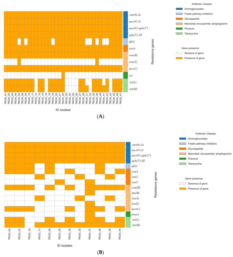
Background/Objectives: Enterococci, particularly Enterococcus faecalis and Enterococcus faecium, are Gram-positive cocci that can cause severe infections in hospitalized patients. The rise of vancomycin-resistant enterococci (VRE) and vancomycin-variable enterococci (VVE) poses significant challenges in healthcare settings due to their resistance to multiple antibiotics. Methods: We conducted a point prevalence survey (PPS) to assess the prevalence of VRE and VVE colonization in hospitalized patients. Rectal swabs were collected from 160 patients and analyzed using molecular assays (MAs) and culture. Whole-genome sequencing (WGS) and core-genome multilocus sequence typing (cgMLST) were performed to identify the genetic diversity. Results: Of the 160 rectal swabs collected, 54 (33.7%) tested positive for the vanA and/or vanB genes. Culture-based methods identified 47 positive samples (29.3%); of these, 44 isolates were identified as E. faecium and 3 as E. faecalis. Based on the resistance profiles, 35 isolates (74.5%) were classified as VRE, while 12 (25.5%) were classified as VVE. WGS and cgMLST analyses identified seven clusters of E. faecium, with sequence type (ST) 80 being the most prevalent. Various resistance genes and virulence factors were identified, and this study also highlighted intra- and inter-ward transmission of VRE strains. Conclusions: Our findings underscore the potential for virulence and resistance of both the VRE and VVE strains, and they highlight the importance of effective infection control measures to prevent their spread. VVE in particular should be carefully monitored as they often escape detection. Integrating molecular data with clinical information will hopefully enhance our ability to predict and prevent future VRE infections.
Keywords: antimicrobial resistance; enterococci; hospital surveillance; vancomycin-resistant; vancomycin-variable; whole-genome sequencing.
Conflict of interest statement
The authors declare no competing interests.
Figures


Similar articles
-
Critical genomic insights into vancomycin-resistant Enterococcus faecium in Lebanon.Microbiol Spectr. 2025 Sep 2;13(9):e0017125. doi: 10.1128/spectrum.00171-25. Epub 2025 Aug 5. Microbiol Spectr. 2025. PMID: 40762484 Free PMC article.
-
Surveillance of vancomycin-resistant enterococci reveals shift in dominating clusters from vanA to vanB Enterococcus faecium clusters, Denmark, 2015 to 2022.Euro Surveill. 2024 Jun;29(23):2300633. doi: 10.2807/1560-7917.ES.2024.29.23.2300633. Euro Surveill. 2024. PMID: 38847117 Free PMC article.
-
VanB transposon analysis detects horizontal gene transfer in vancomycin-resistant Enterococcus faecium: description of two outbreaks.J Hosp Infect. 2025 Aug;162:351-359. doi: 10.1016/j.jhin.2025.04.021. Epub 2025 May 6. J Hosp Infect. 2025. PMID: 40339917
-
Does vancomycin prescribing intervention affect vancomycin-resistant enterococcus infection and colonization in hospitals? A systematic review.BMC Infect Dis. 2007 Apr 10;7:24. doi: 10.1186/1471-2334-7-24. BMC Infect Dis. 2007. PMID: 17425800 Free PMC article.
-
Epidemiology and outcomes of vancomycin-resistant enterococcus infections: a systematic review and meta-analysis.J Hosp Infect. 2023 Nov;141:119-128. doi: 10.1016/j.jhin.2023.09.008. Epub 2023 Sep 19. J Hosp Infect. 2023. PMID: 37734679
References
-
- Global Report on Infection Prevention and Control. 2024. [(accessed on 6 June 2025)]. Available online: https://www.who.int/publications/i/item/9789240103986.
Grants and funding
LinkOut - more resources
Full Text Sources
