

Morphological and Functional Analysis of Residual Lung After Pneumonectomy in Lung Cancer Surgery via 3D-CT Method
- PMID: 40868913
- PMCID: PMC12387389
- DOI: 10.3390/life15081265
Morphological and Functional Analysis of Residual Lung After Pneumonectomy in Lung Cancer Surgery via 3D-CT Method
Abstract
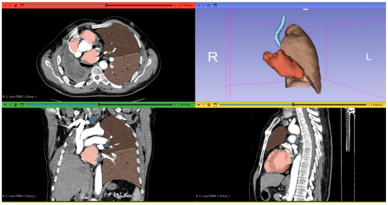
Background: Pneumonectomy is a major surgical option for non-small cell lung cancer (NSCLC). This study evaluates the predictive value of three-dimensional computed tomography (3D-CT)-based lung volume analysis for postoperative function and explores its potential role in preoperative planning, risk assessment, and surgical decision-making. Methods: We evaluated 59 NSCLC patients who underwent pneumonectomy. Pre- and 12-month postoperative spirometry results were compared with anatomical and 3D-CT-based predictions. Residual lung expansion was calculated, and patients were grouped by a 3D-CT-derived volume ratio of ≥1.2 or <1.2. Results: There was a significant correlation between 3D-CT-based predicted FVC and FEV1 and postoperative spirometric values (p < 0.001). The mean residual lung volume expansion ratio was 1.23. Patients with a ratio ≥1.2 had significantly higher postoperative FVC (p = 0.028). Lung expansion was observed in 81.4% of cases. Predicted postoperative FVC (p = 0.023) and FEV1 (p = 0.013) were significantly higher in patients with left pneumonectomy compared to right. Conclusions: 3D-CT-based lung volume calculation reliably predicts postoperative function and matches conventional methods. Contralateral lung expansion positively affects respiratory outcomes. Additionally, 3D-CT analysis supports preoperative planning and risk assessment, contributing to more accurate diagnosis and surgical decisions in NSCLC management.
Keywords: 3D computed tomography (3D-CT); lung volume calculation; pneumonectomy; pulmonary function test; residual lung expansion.
Conflict of interest statement
The authors declare that they have no known competing financial interests or personal relationships that could have appeared to influence the work reported in this paper.
Figures


Similar articles
-
FEV1 and DLCO predicting general complications but not prolonged air leaks in pulmonary segmentectomy.Ther Adv Respir Dis. 2025 Jan-Dec;19:17534666251341777. doi: 10.1177/17534666251341777. Epub 2025 Jul 7. Ther Adv Respir Dis. 2025. PMID: 40624919 Free PMC article.
-
PET-CT for assessing mediastinal lymph node involvement in patients with suspected resectable non-small cell lung cancer.Cochrane Database Syst Rev. 2014 Nov 13;2014(11):CD009519. doi: 10.1002/14651858.CD009519.pub2. Cochrane Database Syst Rev. 2014. PMID: 25393718 Free PMC article.
-
Analysis of Factors Affecting Prolonged Air Leak and Expansion Failure in the Lung after Resection in Patients with Pulmonary Malignancy and Predictive Value of Preoperative Quantitative Chest Computed Tomography.Thorac Cardiovasc Surg. 2025 Aug;73(5):418-426. doi: 10.1055/a-2508-6067. Epub 2025 Jan 21. Thorac Cardiovasc Surg. 2025. PMID: 39837533
-
The role of growth and nutrition in the early origins of spirometric restriction in adult life: a longitudinal, multicohort, population-based study.Lancet Respir Med. 2022 Jan;10(1):59-71. doi: 10.1016/S2213-2600(21)00355-6. Epub 2021 Nov 26. Lancet Respir Med. 2022. PMID: 34843665 Free PMC article.
-
Preoperative exercise training for patients with non-small cell lung cancer.Cochrane Database Syst Rev. 2017 Jun 7;6(6):CD012020. doi: 10.1002/14651858.CD012020.pub2. Cochrane Database Syst Rev. 2017. Update in: Cochrane Database Syst Rev. 2022 Sep 28;9:CD012020. doi: 10.1002/14651858.CD012020.pub3. PMID: 28589547 Free PMC article. Updated.
References
-
- Sharma S., Beshara M., Bora V. Pneumonectomy. [(accessed on 17 January 2025)];Medical Management of the Surgical Patient: A Textbook of Perioperative Medicine [Internet] 2024 :596–597. Available online: https://www.ncbi.nlm.nih.gov/books/NBK555969/
-
- Huang J., Osarogiagbon R.U., Giroux D.J., Nishimura K.K., Bille A., Cardillo G., Detterbeck F., Kernstine K., Kim H.K., Lievens Y., et al. The International Association for the Study of Lung Cancer Staging Project for Lung Cancer: Proposals for the Revision of the N Descriptors in the Forthcoming Ninth Edition of the TNM Classification for Lung Cancer. J. Thorac. Oncol. 2024;19:766–785. doi: 10.1016/j.jtho.2023.10.012. - DOI - PMC - PubMed
Grants and funding
LinkOut - more resources
Full Text Sources
