. 2025 Aug 8;26(16):7697.
doi: 10.3390/ijms26167697.

Microbial Signatures of Obesity-Aggravated Psoriasis: Insights from an Imiquimod-Based Mouse Model

Affiliations

Microbial Signatures of Obesity-Aggravated Psoriasis: Insights from an Imiquimod-Based Mouse Model

Carolina Constantin et al. Int J Mol Sci. .

Abstract

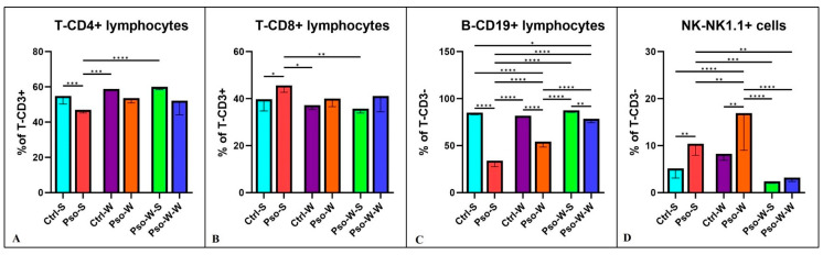
As obesity and Western diet consumption are key factors contributing to gut dysbiosis, we investigated the relationship between intestinal microbiota, obesity, and psoriasis in an imiquimod-based model. C57BL/6 mice were used as follows: psoriasis-induced groups fed continuously with a standard or Western diet, psoriasis-induced group fed with a Western diet and then returned to a standard diet, and controls. For each group, clinicopathological, immune, and metabolic parameters were integrated with microbiome data. The imiquimod-based models displayed human psoriasis features and significant changes in immune parameters. Hence, psoriatic mice on prolonged high-fat intake presented decreased microbial richness and evenness and a gut microbiome composition resembling that of obese mice. Ruminococcus, Clostridium, Desulfovibrio, and Enterorhabdus were the most abundant genera in the obesity-enhanced psoriasis group. Raoultella abundance was linked with psoriasis. Yet, the same pathobionts over-represented in the obese psoriatic mice displayed positive correlations with metabolic stress indicators and proinflammatory factors, indicating potential biomarkers of disease severity. Conversely, Lactobacillus taiwanensis, Alistipes putredinis, and Eubacterium hadrum might be potential taxa for attenuating the metabolic burden in obesity-enhanced psoriasis. Here, we depict the microbial signatures associated with inflammation and metabolic stress in an obesity-aggravated psoriasis mouse model.

Keywords: 16S rRNA sequencing; diet; dysbiosis; microbiota; psoriasis.


Conflict of interest statement

The authors declare no conflicts of interest.

Figures

Figure 1
Figure 1
Comparative evolution of skin inflammatory parameters in Pso-W and Pso-S groups. Daily scoring for erythema (A), skin scaling (B), thickness (C), and PASI score (D) for Pso-W (n = 18) and Pso-S (n = 8) groups; data are presented as mean score ± SD (see Supplementary Material Table S1); n = number of mice. (E) H&E staining of the back skin samples provided from Ctrl-S (E1), Pso-S (E2), Ctrl-W (E3), and Pso-W (E4) groups (scale bar = 400 μm).
Figure 2
Figure 2
Evolution of body weight in experimental groups. (A). Body weight evolution in Ctrl-S and Ctrl-W groups until 14 weeks of age; the results are presented as mean body weight ± SD. (B). Body weight evolution in Ctrl-S, Ctrl-W, Pso-W-S, and Pso-W-W groups until 21 weeks of age; the results are presented as mean values for body weight (see Supplementary Material Table S2).
Figure 3
Figure 3
Splenomegaly assessment. (A) Representative images of spleen harvested from Ctrl-S (A1), Pso-S (A2), Ctrl-W (A3), and Pso-W (A4) mice; (B) weights of the spleens in Ctrl-S vs. Pso-S groups and Ctrl-W vs. Pso-W groups; (C) the SW/BW ratio in Ctrl-S vs. Pso-S groups and Ctrl-W vs. Pso-W groups; the results are presented as mean spleen weight ± SD and mean ratio SW/BW ± SD (see Supplementary Material Table S3). ** p < 0.01; *** p < 0.001.
Figure 4
Figure 4
Lymphocytes’ distribution in peripheral blood. Percentage distribution of T-CD4+ (A), T-CD8+ (B), B-CD19+ (C), and NK-NK1.1+ (D) cells in all experimental groups; the results are presented as percentages of T-CD3ε+ and T-CD3ε lymphocytes (mean ± SD) (see Supplementary Material Table S5). * p < 0.05; ** p < 0.01; *** p < 0.001; **** p < 0.0001.
Figure 5
Figure 5
Serum levels distribution of IL-6 (A), IL-1β (B), IFN-γ (C), and TNF-α (D) in all experimental groups; the results are presented as mean ± SD (see Supplementary Material Table S6). * p < 0.05; ** p < 0.01; *** p < 0.001; **** p < 0.0001.
Figure 6
Figure 6
Serum level distribution of IL-5 (A), IL-10 (B), and IL-17 (C) in all experimental groups; the results are presented as mean ± SD (see Supplementary Material Table S6). * p < 0.05; ** p < 0.01; *** p < 0.001; **** p < 0.0001.
Figure 7
Figure 7
Serum levels distribution of MCP-1 (A), MIP-1α (B), RANTES (C), and PF-4 (D) in all experimental groups; the results are presented as mean ± SD (see Supplementary Material Table S6). * p < 0.05; ** p < 0.01; *** p < 0.001; **** p < 0.0001.
Figure 8
Figure 8
Serum level distribution of ICAM-1 (A), TIMP-1 (B), Leptin (C), and Eotaxin (D) in all experimental groups; the results are presented as mean ± SD (see Supplementary Material Table S6). * p < 0.05; ** p < 0.01; *** p < 0.001; **** p < 0.0001.
Figure 9
Figure 9
Microbiome community composition and diversity analyses: (A) Observed species richness across intervention groups, with leave-one-group-out exclusion strategy shown in facets. Each box represents the alpha diversity distribution within the indicated group after excluding the specified intervention group. (B) Chao1 estimator of species richness for the same leave-one-out exclusion design. Colors indicate experimental groups: Ctrl-S (cyan), Pso-S (coral), Ctrl-W (magenta), Pso-W (orange), Pso-W-S (chartreuse), Pso-W-W (blue). (C) Principal Coordinate Analysis (PCoA) based on Bray–Curtis dissimilarity of microbial communities, showing clustering by intervention group. Ellipses indicate 95% confidence intervals for group centroids. (D) Heatmap of relative abundances, clustered by sample group and taxon. Color scale indicates log-transformed relative abundance. * p < 0.05.
Figure 10
Figure 10
Firmicutes/Bacteroidetes ratio across intervention groups: Log-transformed Firmicutes-to-Bacteroidetes ratios are shown for each experimental group using violin plots overlaid with boxplots. Colors indicate intervention groups: cyan (Ctrl-S), coral (Pso-S), dark magenta (Ctrl-W), orange (Pso-W), chartreuse (Pso-W-S), and blue (Pso-W-W). Wider violins indicate greater density of values. Differences in central tendency and distribution reflect group-specific shifts in relative abundance of these dominant phyla.
Figure 11
Figure 11
Heatmap of Spearman correlations between bacterial genera and host cytokine levels. The heatmap shows Spearman correlation coefficients between the relative abundances of bacterial genera (columns) and circulating cytokine levels (rows). Positive correlations are indicated in red and negative correlations in blue, with color intensity reflecting correlation strength (scale from −1 to +1). Hierarchical clustering of rows and columns highlights patterns of shared associations among bacterial genera and immune markers.
Figure 12
Figure 12
Heatmap of Spearman correlations between bacterial species (species level) and host metabolic parameters. The heatmap displays Spearman correlation coefficients between log-transformed relative abundances of bacterial species (rows, species-level resolution) and host clinical variables (columns): cholesterol, weight, triglycerides, and spleen weight. Positive correlations are shown in red and negative correlations in blue, with color intensity indicating the strength of association. Hierarchical clustering of rows and columns highlights groups of taxa and host traits with similar correlation patterns.
Figure 13
Figure 13
The outline of the experimental model. The graphical scheme includes the main key points of the study and the involved experimental groups (Created in https://BioRender.com).
Figure 14
Figure 14
Schematic E. coli 16S rRNA gene and primer targets. The 16S rRNA gene is about 1542 base pairs long and includes nine hypervariable regions (V1–V9) along with conserved regions. Ion 16S Metagenomics kit contains two sets of primers targeting seven hypervariable regions (V2-4-8 and V3, V6-7, V9) (blue arrows—primers for V2-4-8 regions, purple arrows—primers for V3, V6-7, V9 regions). Created in https://BioRender.com, adapted from ThermoFisher Scientific, Inc. [83].

References

    1. Turnbaugh P.J., Bäckhed F., Fulton L., Gordon J.I. Diet-induced obesity is linked to marked but reversible alterations in the mouse distal gut microbiome. Cell Host Microbe. 2008;3:213–223. doi: 10.1016/j.chom.2008.02.015. - DOI - PMC - PubMed
    1. Sun C., Chen L., Yang H., Sun H., Xie Z., Zhao B., Jiang X., Qin B., Shen Z. Involvement of Gut Microbiota in the Development of Psoriasis Vulgaris. Front. Nutr. 2021;8:761978. doi: 10.3389/fnut.2021.761978. - DOI - PMC - PubMed
    1. Macpherson A.J., Harris N.L. Interactions between commensal intestinal bacteria and the immune system. Nat. Rev. Immunol. 2004;4:478–485. doi: 10.1038/nri1373. - DOI - PubMed
    1. Derrien M., Belzer C., de Vos W.M. Akkermansia muciniphila and its role in regulating host functions. Microb. Pathog. 2017;106:171–181. doi: 10.1016/j.micpath.2016.02.005. - DOI - PubMed
    1. Everard A., Belzer C., Geurts L., Ouwerkerk J.P., Druart C., Bindels L.B., Guiot Y., Derrien M., Muccioli G.G., Delzenne N.M., et al. Cross-talk between Akkermansia muciniphila and intestinal epithelium controls diet-induced obesity. Proc. Natl. Acad. Sci. USA. 2013;110:9066–9071. doi: 10.1073/pnas.1219451110. - DOI - PMC - PubMed

LinkOut - more resources