. 2025 Aug 12;26(16):7781.
doi: 10.3390/ijms26167781.

Porphyromonas gingivalis GroEL Accelerates Abdominal Aortic Aneurysm Formation by Induction of M1 Polarization in Macrophages

Affiliations

Porphyromonas gingivalis GroEL Accelerates Abdominal Aortic Aneurysm Formation by Induction of M1 Polarization in Macrophages

Yi-Wen Lin et al. Int J Mol Sci. .

Abstract

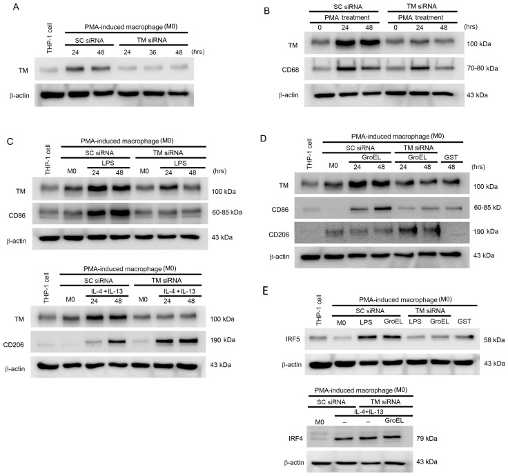
Abdominal aortic aneurysm (AAA) is a life-threatening vascular disease characterized by chronic inflammation, extracellular matrix degradation, and smooth muscle cell apoptosis. Porphyromonas gingivalis (P. gingivalis), a key periodontal pathogen, has been implicated in the progression of cardiovascular diseases, including AAA, but the underlying mechanisms remain unclear. In this study, we investigated the role of GroEL, a bacterial heat shock protein 60 homolog derived from P. gingivalis, in AAA development. We employed a CaCl2-induced AAA mouse model to evaluate the in vivo effects of GroEL. Mice received periaortic CaCl2 application followed by intravenous injections of recombinant GroEL. Histological analyses were performed to assess aneurysmal dilation, elastin degradation, and inflammatory cell infiltration. Flow cytometry and immunohistochemistry were used to determine macrophage phenotypes, while cytokine profiles were quantified via ELISA. In vitro, THP-1 monocytes were treated with GroEL to evaluate its impact on macrophage polarization and cytokine expression. Our results showed that GroEL administration significantly enhanced aortic diameter expansion and elastin breakdown, accompanied by increased infiltration of M1-like macrophages and elevated levels of pro-inflammatory cytokines such as TNF-α and IL-6. In vitro findings confirmed that GroEL promotes M1 polarization and inhibits M2 marker expression in THP-1-derived macrophages. These findings suggest that P. gingivalis-derived GroEL plays a pathogenic role in AAA by modulating macrophage polarization toward a pro-inflammatory phenotype. Targeting microbial components such as GroEL may offer new therapeutic strategies for AAA management.

Keywords: GroEL; IRF5; P. gingivalis; abdominal aortic aneurysm; macrophages; thrombomodulin.


Conflict of interest statement

The authors declare no conflicts of interest.

Figures

Figure 1
Figure 1
GroEL accelerates AAA formation in CaCl2 immersion-induced mice. (A) After the experiment, the total aortas of the rats were removed, and images were captured using a digital camera (left panel/green background). The black arrows indicate aneurysm lesions. The dissected abdominal aortas were then analyzed using micro-CT. Representative images of the tissues from each group are presented in the right panel (black background). The white arrows indicate vascular calcification and aneurysm lesions. (B) Graph presenting the AAA incidence of each group. (C) The upper column shows the abdominal aortas from rats stained with H&E. The images are 100× magnified. The arrowheads indicate aneurysm lesions. The lower column presents the integrity of the elastic fibers of abdominal aorta cross-sections observed using VVG staining. The images are 400× magnified. (D,E) Graphs displaying the quantification of the luminal diameter (D) and total length of elastic fibers (E). All the results in the graphs are expressed as the mean ± SD. * Statistical significance was set at p < 0.05.
Figure 2
Figure 2
GroEL induces protein expression associated with inflammation and AAA formation in THP-1 cells. THP-1 cells were stimulated for 24–72 h with 10 μg/mL of GroEL. Treatment with 10 μg/mL of GST for 72 h served as a negative control. (A) Images showing the western blot analyses of TLR4, MMP-2, and phosphorylated iNOS proteins. (B) Images showing western blot analyses of PCNA, p21, ICAM-1, and CD18/integrin β2. The expression levels of total β-actin and total iNOS were used as loading controls.
Figure 3
Figure 3
GroEL induces M1 macrophage polarization and inhibits M2 macrophage polarization. THP-1 cells were stimulated with 10 μM PMA for 24 h to differentiate into attached cells (M0 macrophages). (A) The left panel shows M0 macrophages stimulated with LPS. The middle panel shows M0 macrophages stimulated with GroEL. The right panel shows M0 macrophages stimulated with LPS first and GroEL afterwards. Total proteins were extracted, and western blot analysis of iNOS was performed. (B) The left panel shows M0 macrophages stimulated with 25 ng/mL IL-4 plus 25 ng/mL IL-13. The middle panel shows M0 macrophages stimulated with GroEL. The right panel shows M0 macrophages stimulated with IL-4 plus IL-13 first and GroEL afterwards. Total proteins were extracted, and western blot analysis of arginase-1 was performed. (C) The upper panel shows M0 macrophages stimulated with LPS or GroEL. The lower panel shows M0 macrophages stimulated with LPS with or without GroEL. Total proteins were extracted, and western blot analysis of CD86 was performed. (D) The upper panel shows M0 macrophages stimulated with 25 ng/mL IL-4 and 25 ng/mL IL-13 or GroEL; the lower panel shows M0 macrophages stimulated with IL-4 and IL-13 with or without GroEL. Total proteins were extracted, and western blot analysis of CD206 was performed. Treatment with 10 μg/mL GST was used to exclude the effect of the tagged protein on the recombinant GroEL protein. The expression of total β-actin was used as a loading control.
Figure 4
Figure 4
GroEL induces M1 macrophage polarization via TM and IRF5 expression. (A) THP-1 cells were stimulated with 10 μM PMA for 24 h to differentiate into attached cells (M0 macrophages), then cells were transfected with TM siRNA for 24–48 h, and the TM in total cell lysate was analyzed using western blot analysis. The scramble siRNA (SC siRNA) was used as a control. (B) THP-1 cells were transfected with or without TM siRNA, followed by 10 μM PMA treatment for 24–48 h. (C) THP-1 cells were stimulated with 10 μM PMA for 24 h to differentiate into M0 macrophages. The upper panel shows M0 macrophages transfected with or without TM siRNA, followed by 1 μg/mL LPS treatment for 24–48 h. The lower panel shows M0 macrophages transfected with or without TM siRNA, followed by 25 ng/mL IL-4 plus 25 ng/mL IL-13 treatment for 24–48 h. The total proteins were extracted, and western blot analyses of TM, CD68, CD86, and CD206 expressions were performed. (D) M0 macrophages were transfected with or without TM siRNA, followed by 10 μg/mL GroEL treatment for 24–48 h. (E) The upper panel shows M0 macrophages transfected with or without TM siRNA, followed by 1 μg/mL LPS or 10 μg/mL GroEL treatment for 48 h. The lower panel shows M0 macrophages transfected with or without TM siRNA, followed by 25 ng/mL IL-4 plus 25 ng/mL IL-13 combined with or without 10 μg/mL GroEL treatment. The total proteins were extracted, and western blot analyses of IRF5 or IRF4 expressions were performed. Treatment with 10 μg/mL GST for 48 h was used to exclude the effect of the tag protein on the recombinant GroEL protein. The expression of total β-actin was used as a loading control in this study.
Figure 5
Figure 5
GroEL aggravates the occurrence of AAA, promotes infiltration of M1 macrophages, and inhibits polarization of M2 macrophages in the vessel walls in Ang II-induced mice. Images show aortic sections stained using (A) rabbit anti-TM Ab, (B) rabbit anti-CD11c Ab, (C) rabbit anti-CD206 Ab, and (D) isotype control rabbit IgG. In the upper panel, black arrows and arrow heads indicate aneurysm lesions in 80× magnification. The lower panel presents the boxes in the upper panels magnified at 200×. Bar graphs display the TM-expressed cells, CD11c-positive cells, and CD206-positive cells infiltrating the aneurysm lesion, counted under a high-power field (200×). All results in the graphs are expressed as the mean ± SD. * Statistical significance was set at p < 0.05.

Similar articles

References

    1. Ding F., Wu D., Han X., Cheng L.J., Sun Z., Lv Y.L. Oral hygiene and periodontal conditions in the Chinese patients with aortic aneurysm. BMC Oral Health. 2018;18:136. doi: 10.1186/s12903-018-0594-3. - DOI - PMC - PubMed
    1. Suzuki J., Aoyama N., Aoki M., Tada Y., Wakayama K., Akazawa H., Shigematsu K., Hoshina K., Izumi Y., Komuro I., et al. High incidence of periodontitis in Japanese patients with abdominal aortic aneurysm. Int. Heart J. 2014;55:268–270. doi: 10.1536/ihj.13-301. - DOI - PubMed
    1. Suzuki J., Aoyama N., Aoki M., Tada Y., Wakayama K., Akazawa H., Shigematsu K., Hoshina K., Izumi Y., Komuro I., et al. Incidence of periodontitis in Japanese patients with cardiovascular diseases: A comparison between abdominal aortic aneurysm and arrhythmia. Heart Vessel. 2015;30:498–502. doi: 10.1007/s00380-014-0507-6. - DOI - PubMed
    1. Bi C., Liu B., Gao P., Wang C., Fang S., Huo Z., Song Q., Dong D., Wu X., Li G. RAGE deficiency ameliorates abdominal aortic aneurysm progression. Inflamm. Res. 2025;74:63. doi: 10.1007/s00011-025-02027-2. - DOI - PubMed
    1. Kurihara N., Inoue Y., Iwai T., Umeda M., Huang Y., Ishikawa I. Detection and localization of periodontopathic bacteria in abdominal aortic aneurysms. Eur. J. Vasc. Endovasc. Surg. 2004;28:553–558. doi: 10.1016/j.ejvs.2004.08.010. - DOI - PubMed

LinkOut - more resources