. 2025 Aug 13;26(16):7810.
doi: 10.3390/ijms26167810.

Integrative High-Throughput RNAi Screening Identifies BRSK1, STK32C and STK40 as Novel Activators of YAP/TAZ

Affiliations

Integrative High-Throughput RNAi Screening Identifies BRSK1, STK32C and STK40 as Novel Activators of YAP/TAZ

Mandeep K Gill et al. Int J Mol Sci. .

Abstract

Disruption of the Hippo pathway leads to activation of the YAP/TAZ transcriptional program which promotes tumor initiation, progression and metastasis in diverse cancers. Aggressive triple-negative breast cancers (TNBC) lack an effective therapy; thus, inactivating YAP and TAZ has emerged as an attractive approach and a new treatment modality. Thus, we performed two complementary high-throughput RNAi-based kinome screens to uncover cancer-associated activators of YAP/TAZ in two TNBC cell lines, MDA-MB231 and MDA-MB468. Integrated analysis that combined a YAP/TAZ localization screen with a TEAD-luciferase reporter screen, identified novel regulators including BRSK1, STK32C and STK40. The AMPK family members NUAKs, MARKs and SIKs are known to inhibit the Hippo kinase cassette; here, we uncover BRSK1, another AMPK family member as a regulator of YAP/TAZ. We also reveal that two poorly studied kinases, STK32C, a member of the AGC family, and STK40, a pseudokinase, can also inhibit the activity of YAP/TAZ. Thus, our studies expand the repertoire of known AMPK family members and reveal two new kinases that modulate the Hippo pathway and may play a role in YAP/TAZ driven breast cancers. Further analysis of other screen hits may similarly uncover new regulators that could be targeted for therapeutic interventions.

Keywords: AMPK family members; BRSK1; Hippo pathway; RNAi screening; STK32C; STK40; YAP/TAZ; triple-negative breast cancer.


Conflict of interest statement

The authors declare no conflicts of interest.

Figures

Figure 1
Figure 1
Analysis of high-throughput RNAi kinome screen controls and selected cell lines. (a) A schematic depicting the siRNA kinome screen. (b) Predominantly nuclear localization of YAP/TAZ in selected TNBC cell lines. Representative images from MDA-MB231 and MDA-MB468 cells showing localization of endogenous YAP/TAZ (green) with nuclei co-stained with DAPI (blue) were obtained by immunofluorescence confocal microscopy. Scale bar, 25 µm. (c) Loss of YAP suppresses wild-type (WT) but not mutant (MUT) TEAD-luciferase reporter activity in MDA-MB231 and MDA-MB468 cells. (siControl:−, siYAP:+). Data is plotted as the mean +/− SD (n = 3). (d) Loss of MARK4 promotes cytoplasmic localization of YAP/TAZ. Representative images from the screen collected by the IN Cell Analyzer 6000 (GE Healthcare, Chicago, IL, USA) are shown. Scale bar, 25 µm (e) The frequency distribution of controls combined from two independent runs is depicted as a histogram, where the number of samples out of a total of 144 siCtl and 36 siMARK4 at the given Z-score (determined on the basis of the entire screen) in each bin is plotted. Controls and siMARK4 samples are marked with green and blue bars, respectively.
Figure 2
Figure 2
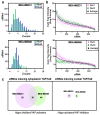
Analysis of high-throughput YAP/TAZ localization screen. (a) The frequency distribution of siRNAs is depicted as a histogram, where the number of siRNAs in each bin is plotted. (b) Screen results are rank-ordered by the average Z-score. (c) Venn diagrams indicating the number of candidate hits defined by the localization screens in the two cell lines.
Figure 3
Figure 3
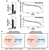
Analysis of high-throughput TEAD-reporter screen. (a) Loss of YAP reduces TEAD-reporter activity. Data is plotted as the mean ± SEM. (b) Screen results are rank-ordered by fold change. (c) Venn diagrams indicating the number of candidate hits defined by the TEAD-reporter screens in the two cell lines.
Figure 4
Figure 4
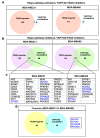
Identification of screen hits. (a,b) Venn diagram indicating the number of candidate hits defined by the two parallel screens. (c) Identified hits common to both localization and TEAD-reporter screens for each line are listed. The four common genes are highlighted in blue text. (d) Identified hits common to both localization and TEAD-reporter screens in both cell lines with 4 common genes listed.
Figure 5
Figure 5
BRSK1 regulates YAP/TAZ localization, transcriptional activity and growth in MDA-MB231 cells. (a) Loss of BRSK1 expression promotes cytoplasmic localization of YAP/TAZ. Representative immunofluorescence image of endogenous YAP/TAZ (green) with nuclei co-stained with DAPI (blue) visualized by confocal microscopy in MDA-MB231 cells, transfected with control (−) or a pool of siRNAs targeting BRSK1 (left). The percentage of cells with equivalent nuclear and cytoplasmic (N + C), predominantly nuclear (N>C) or predominantly cytoplasmic (C>N) YAP/TAZ localization is plotted (right). Scale bar, 25 µm. (b) Loss of BRSK1 expression decreases TEAD-luciferase reporter activity. YAP/TAZ transcriptional activity was assessed using a wild-type (WT) and mutant (MUT) TEAD-luciferase reporter in MDA-MB231 cells, transfected with control (−) or a pool of siRNAs targeting BRSK1. Data is plotted relative to siCTL, and the mean ± SD of three independent experiments is shown. (ce) Loss of BRSK1 expression decreases YAP/TAZ target gene expression. Relative expression of ANKRD1, CTGF, CYR61 and knockdown efficiency of BRSK1, transfected as a pool or as individual (numbered 1 and 2) siRNAs, as measured by qPCR in MDA-MB231 (c,d) or MDA-MB468 cells (e) is plotted. The mean ± SD of three independent experiments is shown. (f) Loss of BRSK1 inhibits the growth of MDA-MB231 cells as measured by DAPI staining. Data is plotted as the mean ± SD of a representative experiment, n = 3.
Figure 6
Figure 6
Overexpression of AMPK-related family members in breast cancer patient samples. An oncoprint generated in cBioportal depicts individual samples displaying overexpression (>2 standard deviations above the mean compared to diploids) of the indicated AMPK-related family members, known or identified in this study to act as YAP/TAZ activators and for YAP and TAZ. The plot was cropped to remove unaltered samples.
Figure 7
Figure 7
STK32C and STK40 regulates YAP/TAZ localization, transcriptional activity and growth in MDA-MB231. (a) Loss of STK32C or STK40 expression promotes cytoplasmic localization of YAP/TAZ. Representative immunofluorescence image of YAP/TAZ (green) with nuclei co-stained with DAPI (blue) in MDA-MB231 cells, transfected with control (−) or a pool of siRNAs, targeting STK32C or STK40 (left). The percentage of cells with equivalent nuclear and cytoplasmic (N + C), predominantly nuclear (N>C) or predominantly cytoplasmic (C>N) YAP/TAZ localization is plotted (right). Scale bar, 25 µm. (b,c) Loss of STK32C or (d,e) STK40 expression in MDA-MB231 cells, transfected with control (−), pool or individual (numbered 1 or 2) siRNAs decreases YAP/TAZ target gene expression. Relative expression of ANKRD1, CTGF, CYR61 and knockdown efficiency of STK32C or STK40 as measured by qPCR is plotted. The mean ± SD of two independent experiments is shown. (f,g) Loss of STK32C or STK40 using siRNA pools (+) inhibits the growth of MDA-MB231 cells as measured by DAPI staining. Data is plotted as the mean ± SD of a representative experiment, n = 3. STK32C or STK40 knockdown efficiency (right) is plotted as mean ± range of a representative experiment.

References

    1. Kim J., Harper A., McCormack V., Sung H., Houssami N., Morgan E., Mutebi M., Garvey G., Soerjomataram I., Fidler-Benaoudia M.M. Global patterns and trends in breast cancer incidence and mortality across 185 countries. Nat. Med. 2025;31:1154–1162. doi: 10.1038/s41591-025-03502-3. - DOI - PubMed
    1. Harbeck N., Penault-Llorca F., Cortes J., Gnant M., Houssami N., Poortmans P., Ruddy K., Tsang J., Cardoso F. Breast cancer. Nat. Rev. Dis. Primers. 2019;5:66. doi: 10.1038/s41572-019-0111-2. - DOI - PubMed
    1. Perou C.M., Sorlie T., Eisen M.B., van de Rijn M., Jeffrey S.S., Rees C.A., Pollack J.R., Ross D.T., Johnsen H., Akslen L.A., et al. Molecular portraits of human breast tumours. Nature. 2000;406:747–752. doi: 10.1038/35021093. - DOI - PubMed
    1. Vagia E., Mahalingam D., Cristofanilli M. The Landscape of Targeted Therapies in TNBC. Cancers. 2020;12:916. doi: 10.3390/cancers12040916. - DOI - PMC - PubMed
    1. Manning S.A., Kroeger B., Harvey K.F. The regulation of Yorkie, YAP and TAZ: New insights into the Hippo pathway. Development. 2020;147:dev179069. doi: 10.1242/dev.179069. - DOI - PubMed

MeSH terms

Substances

LinkOut - more resources