. 2025 Aug 15;26(16):7889.
doi: 10.3390/ijms26167889.

MicroRNAs and Their Inhibition in Modulating SLC5A8 Expression in the Context of Papillary Thyroid Carcinoma

Affiliations

MicroRNAs and Their Inhibition in Modulating SLC5A8 Expression in the Context of Papillary Thyroid Carcinoma

Wojciech Gierlikowski et al. Int J Mol Sci. .

Abstract

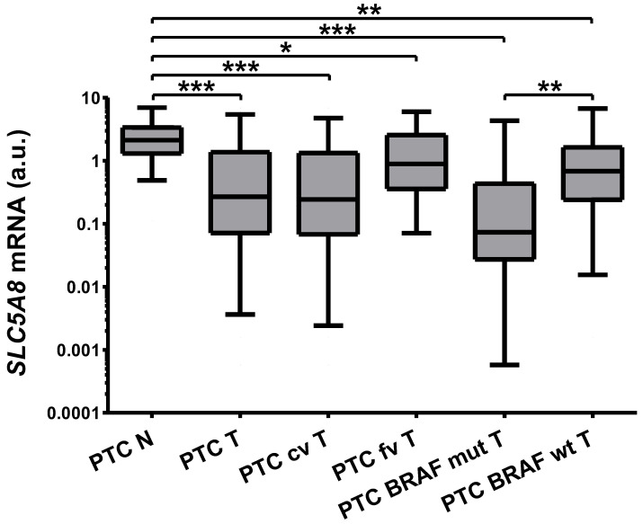
SLC5A8 is a protein coded by the SLC5A8 gene, and has been proposed as a tumor suppressor and iodide transporter. Its expression is reduced in papillary thyroid carcinoma (PTC), yet the mechanisms underlying this phenomenon are largely unknown. We hypothesized that SLC5A8 expression in PTC is reduced by microRNAs and can be modulated by their inhibition. We used real-time PCR to analyze the expression of SLC5A8 and the microRNAs of interest in a set of 49 PTC/normal tissue pairs. We used an in silico approach to identify microRNAs upregulated in PTC and putatively binding to the SLC5A8 transcript. Luciferase assays were performed to confirm the direct binding of synthetic microRNAs to the 3'UTR of SLC5A8. Subsequently, using mir-expressing plasmids and microRNA sponges, including a microRNA sponge designed to simultaneously inhibit three selected microRNAs, we checked the impact of the modulation of microRNAs on endogenous SLC5A8. Finally, we investigated if modulation of SLC5A8 induces changes in transcriptomes. We confirmed the downregulation of SLC5A8 in PTC. In silico analysis revealed microRNAs potentially targeting SLC5A8. Luciferase assay confirmed direct binding between the 3'UTR of SLC5A8 and miR-181a-5p, miR-182-5p, and miR-494-3p. MiR-181a-5p and miR-182-5p were upregulated in PTC. In HEK293 cell lines, transfection with mir-181a- and mir-182-expressing plasmids decreased endogenous SLC5A8 mRNA, while silencing of miR-181a-5p, miR-182-5p, miR-494-3p, and all three microRNAs simultaneously increased SLC5A8 expression; however, only simultaneous inhibition was able to induce changes visible for SLC5A8 protein. Changes in SLC5A8 expression did not alter the whole transcriptome significantly. This study shows microRNA-dependent regulation of SLC5A8 expression and underlines the potential effectiveness of simultaneous inhibition of a few microRNAs to derepress their common target.

Keywords: AIT; PTC; SLC5A8; microRNA; papillary thyroid carcinoma; thyroid carcinoma.


Conflict of interest statement

The authors declare no conflicts of interest. The funders had no role in the design of the study; in the collection, analyses, or interpretation of data; in the writing of the manuscript; or in the decision to publish the results.

Figures

Figure 1
Figure 1
The expression of SLC5A8 is 7.87-fold (p < 0.001) reduced in PTC (PTC T, n = 49) compared with normal adjacent thyroid tissue obtained simultaneously from the contralateral lobe of the same patient (PTC N, n = 49). The reduction was more profound in BRAF-mutated (PTC BRAF mut T, n = 19) than in the wild-type BRAF group (PTC BRAF wt T, n = 23, 9-fold, p = 0.0047) and in the classic variant of PTC (PTC cv T, n = 43) compared with the follicular variant (PTC fv T, n = 6). Expression in all healthy tissue is plotted as it includes the other control subset, which statistically do not differ. The graph shows the expression of SLC5A8 in thyroid tissue samples normalized against HPRT. Data are expressed as median, interquartile range, and a 5–95 percentile. A logarithmic scale was used. Statistical analysis was performed with a Wilcoxon matched-pairs signed-ranks test and Mann–Whitney U test (the latter to compare SLC5A8 expression in tumor tissue depending on BRAF status. * p < 0.05, ** p < 0.01, *** p < 0.001).
Figure 2
Figure 2
Putative binding sites of selected microRNAs: miR-29a-3p (A,B), miR-92b-3p (C), miR-181a-5p (D), miR-182-5p (E), and miR-494-3p (F) in SLC5A8 3′UTR according to in silico analysis. Numbers in the bottom-left corner of each panel represent the position within the 3′UTR sequence (microRNA.org).
Figure 3
Figure 3
Binding of microRNAs with 3′UTR of SLC5A8 using luciferase reporter assay. Luciferase activity is reduced upon transfection with miR-181a-5p (by 12%, p = 0.04), miR-182-5p (by 23%, p = 0.03), and miR-494-3p (by 15%, p = 0.007). Luciferase activity is shown as a percentage relative to the control (cells transfected with a scrambled control microRNA). The graph shows the mean along with deviations from mean (SEM). Statistical analysis was performed using an unpaired t test (* p < 0.05, ** p < 0.01).
Figure 4
Figure 4
The ratio of expression of the analyzed microRNAs in a tumor (PTC-T) compared to normal adjacent thyroid tissue obtained simultaneously from the contralateral lobe of the same patient (PTC-N, N = 49). The mean expression difference was 1.22 for miR-181a-5p (p = 0.0007) and 1.38-fold (p = 0.002) for miR-182-5p. Data are expressed as median, interquartile range, and 10–90 percentile. Statistical analysis was performed with a Wilcoxon matched-pairs signed-ranks test to compare expression of each microRNA in PTC-T vs. PTC-N tissue (** p < 0.01, *** p < 0.001). A logarithmic scale was used.
Figure 5
Figure 5
Verification of plasmids functionality. (a) Transfection of HEK293 cells with SCL5A8-expressing plasmid led to 105-fold increase in its expression (p = 0.004). The results were normalized against HPRT expression. (b) Expression of microRNAs upon the transfection with a microRNA-expressing plasmids. Transfection of HeLa cells with mir-181a, -182, and -494 expressing plasmids led to a 32-, 50-, and 229-fold increase in their expression, respectively (all p < 0.001). The results were normalized to U44 expression. (c) As sponge is cloned downstream of the luciferase gene, the activity of luciferase expressed from the sponge plasmids is reduced upon transfection with a corresponding microRNA. Luminescence mediated by the sponge ctrl is reduced upon co-transfection with the mir–ctrl-expressing plasmid in comparison to co-transfection with a mix of mir-181a-, mir-182-, and mir-494-expressing plasmids by 8% (p = 0.045). Similarly, co-transfection with sponge-181 and mir-181-expressing plasmids led to reduction in luciferase activity by 22% (p = 0.012), whilst for sponge-182 and mir-182 it was by 25% (p < 0.0001), and for sponge-494 and mir-494 it was by 21% (p = 0.003). (d) Sponge–mix plasmid was designed to bind with each of miR-181a-5p, miR-182, and miR-494-3p. Co-transfection of HeLa cells with the sponge–mix plasmid with microRNA-expressing plasmids led to decrease in luciferase activity by 45% in the case of mir-181a, 45% for mir-182, and 31% for mir-494 (all p < 0.001). The results were normalized to Renilla luciferase. The graphs show the mean along with deviations from mean (SEM). Statistical analysis was performed using an unpaired t test (* p < 0.05, ** p < 0.01, *** p < 0.001). A logarithmic scale was used in (a,b).
Figure 6
Figure 6
Expression of endogenous SLC5A8 in the HEK293 cell line transfected with microRNA- or sponge-expressing plasmids. (a) Transfection with mir-181a and mir-182 resulted in a reduction in SLC5A8 mRNA measured in real-time PCR assay by 33% (p = 0.003) and 32% (p = 0.01), respectively. (b) The effect of microRNAs overexpression was not visible on the protein level. (c) Silencing of miR-181a-5p increased SLC5A8 mRNA expression 2-fold (p = 0.004), for miR-182-5p it was 6-fold (p = 0.001), and for miR-494-3p it was 7-fold (p = 0.007), whereas simultaneous inhibition of these microRNAs by sponge–mix increased SLC5A8 expression 2-fold (p = 0.006). (d) SLC5A8 protein expression was increased by 28% (p = 0.002) only upon simultaneous inhibition of the microRNAs by the sponge–mix, whereas the differences in expression of SLC5A8 upon transfection with the other sponges are not significant. Data are expressed as mean values +/− SEM (a,d) or median +/− interquartile range (b,c). Statistical analysis was performed using an unpaired t test (a,d) or a Mann–Whitney test ((b,c) * p < 0.05, ** p < 0.01, *** p < 0.001). A logarithmic scale in (c) was used.

Similar articles

References

    1. Ganapathy V., Gopal E., Miyauchi S., Prasad P.D. Biological functions of SLC5A8, a candidate tumour suppressor. Biochem. Soc. Trans. 2005;33:237–240. doi: 10.1042/BST0330237. - DOI - PubMed
    1. Rodriguez A., Perron B., Lacroix L., Caillou B., Leblanc G., Schlumberger M., Bidart J., Pourcher T. Identification and characterization of a putative human iodide transporter located at the apical membrane of thyrocytes. J. Clin. Endocrinol. Metab. 2002;87:3500–3503. doi: 10.1210/jcem.87.7.8797. - DOI - PubMed
    1. Li H., Myeroff L., Smiraglia D., Romero M., Pretlow T., Kasturi L., Lutterbaugh J., Rerko R., Casey G., Issa J., et al. SLC5A8, a sodium transporter, is a tumor suppressor gene silenced by methylation in human colon aberrant crypt foci and cancers. Proc. Natl. Acad. Sci. USA. 2003;100:8412–8417. doi: 10.1073/pnas.1430846100. - DOI - PMC - PubMed
    1. Hong C., Maunakea A., Jun P., Bollen A.W., Hodgson J.G., Goldenberg D.D., Weiss W.A., Costello J.F. Shared epigenetic mechanisms in human and mouse gliomas inactivate expression of the growth suppressor SLC5A8. Cancer Res. 2005;65:3617–3623. doi: 10.1158/0008-5472.CAN-05-0048. - DOI - PubMed
    1. Park J.Y., Helm J.F., Zheng W., Ly Q.P., Hodul P.J., Centeno B.A., Malafa M.P. Silencing of the candidate tumor suppressor gene solute carrier family 5 member 8 (SLC5A8) in human pancreatic cancer. Pancreas. 2008;36:e32–e39. doi: 10.1097/MPA.0b013e3181630ffe. - DOI - PubMed

LinkOut - more resources